Sunday, 29 March 2015

Extraction, amplification and magnification in the Life Sciences


The final lab sessions of this term, that form the core of the UTC’s skills passport, involves the technique called simply, PCR: the Polymerase Chain Reaction. It is arguably the most important laboratory method to come out of Molecular Biology apart from nucleotide sequencing, although I am sure some would disagree! Nevertheless, since the concept was proposed by the distinguished Biochemist, Arthur Kornberg and subsequently developed into the practical technique we know today by Kary Mullis, it has become invaluable not only to Life Science Researchers, but to the Police, Medics, Archaeologists, Historians and Lawyers! Before I discuss the methodology in detail, there are several generic principles that underpin the PCR method, and the technique of PCR provides me with an opportunity to discuss them. The first is the phenomenon that is perhaps best described in large scale data analysis using computational methods: "garbage in garbage out", a phrase attributed in more eloquent form to the computational pioneer Charles Babbage:

On two occasions I have been asked, "Pray, Mr. Babbage, if you put into the machine wrong figures, will the right answers come out?" ... I am not able rightly to apprehend the kind of confusion of ideas that could provoke such a question.
Image result for tooth extractionPersonally, I don't believe that enough experimental scientists appreciate the significance of sample extraction and preparation. After all, we place a great deal of trust in our dentist if an extraction is required! This is largely an experiential view and one that I was forced to confront when I first had to come to terms with transitioning basic to applied research in collaboration with a number of commercial organisations. The robustness of a method, such as ion exchange chromatography, electron microscopy, NMR spectroscopy etc., all stand or fall on the quality and quantity of the input sample. It doesn't matter how sophisticated your instrument is, if the sample under investigation does not meet a certain level of purity, or is present at too low a concentration, or has been prepared at the wrong pH, or salt concentration etc., you may as well not bother! As Arthur Kornberg himself commented: "Why waste pure thoughts on impure proteins!"

If part of your business is the sale of DNA purification kits, then you will not only sell on price, but on the simplicity, efficiency and reproducibility of your particular kit in producing samples fit for purpose in the downstream process. In most research labs, a method is often developed by an individual, which may subsequently becomes a key part of that particular laboratory's repertoire (often for several years). This process of method development and dissemination to the wider scientific community has been a pillar of experimental science in Universities and Research Institutes for many years (this is reflected in the many journals and books dedicated to experimental methodology). However, in our high-throughput, data-hungry world, the importance of personal experimental failure and improvement in the laboratory, has largely been sacrificed in the oncoming juggernaut which is our thirst for answers. Clearly, the current search for an effective Ebola vaccine, in the same way that Jonas Salk raced to deliver his polio vaccine, against a background of escalating human tragedy, focuses the mind in an important way. In many ways, the two competing forces: delivery of solutions and the provision of high quality scientific training are complementary: a good scientist knows when to slow down and take care at the bench, and when to buy an off the peg solution to move a project along. What I would advocate is that greater importance is placed on teaching robust sample preparation methods, as a means of mitigating some of the trouble-shooting shortcomings of many young scientists, in order that they are capable of making informed decisions in their work. The quality and quantity of the input sample in PCR (usually referred to as the "template" is critical for success.
Moving on, amplification and magnification are generally associated with electronics and optics respectively. However, they both have the same general meaning. Consider a singer in a small room ( a typical classroom for example): the singer would normally be audible to everyone in that room. As the size of the room increases, for example a small assembly hall or a large theatre, the singer becomes inaudible, especially at the back of the room. Clearly this phenomenon limited the size of many concert venues, and at the same time stimulated architects to incorporate acoustic criteria into their designs. Nevertheless, there is always a point reached where the singer becomes inaudible to even someone with the keenest of ears! This not only stimulated architects, but also electronic engineers, and thus was born, the microphone: a device for capturing sound waves and converting them into an electrical current. Similarly an amplifier is an electronic device that increases the power of a signal, such as "amplifying" the normal level of sound from a solid body electric guitar. As a result of these two developments, it has become commonplace to hold concerts in football stadia, with suitable equipment. In an analogous way, it was impossible to observe single bacterial cells and certainly not viruses, until the development of optical and electron microscopes respectively. In the former, usually through a combination of optical lenses, the image of a small object can be obtained and otherwise "invisible" objects appear as "virtual" images. Electron microscopes use electrons as the source of illumination and can produce images around 5 000 times greater in magnification, owing to the difference in resolving power resulting in the use of electrons. PCR is similarly a method that does for DNA samples what the electron microscope does for imaging, or the modern microphone does for sound. So let's consider how PCR works.

The objective of (the) PCR is to amplify a specific sequence or set of sequences from a vanishingly small sample of DNA (it could be a research sample or a scene of crime swab). The products of this amplification are called amplicons, and the sample of DNA is referred to as the template. The components of PCRs are pretty logical: a DNA Polymerase enzyme to replicate the DNA, a mixture of dATP, dGTP, dCTP and dTTP (collectively called dNTPs); the building blocks of DNA. All polymerases usually require magnesium ions and a suitable buffer. The missing ingredients are the "primers". These are short (usually between 18-50 nucleotides in length) sequences of DNA; they must be complementary to the ends of the amplicon, following the standard Watson-Crick base pairing rules (A pairs with T and G with C). It is important to appreciate that the two strands of the double helix are anti-parallel. This is illustrated in the top RHS figure. We refer to the direction of the strand as 5'-3' (spoken 5-prime to 3-prime), this is reflected in the orientation of the sugar phosphates that form the backbone of the DNA molecule.

The key to amplification lies in a series of repeated denaturation steps, or cycles during which the Watson-Crick base pairs are "broken", allowing the primers to find their complementary sites. Denaturation is achieved by heating the reaction; the primers (which are in considerable excess over the template DNA) then anneal to form the substrate binding site for the polymerase enzyme. The dNTPs are then incorporated as the polymerase copies each strand in the 5'-3' direction. DNA replication proceeds only in one direction, each double helix that is copied is analogous to two railway lines (say from Liverpool to London): just as the train keeps to one set of tracks, so to do the DNA polymerase molecules. During replication in vivo, this unidirectionality causes topological challenges for the genome in the cell, but this will be the subject of another post.

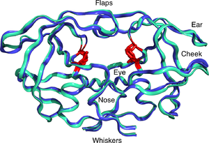
By heating and cooling around 20-30 times, the amplification proceeds as 1 duplex becomes 2, 2 become 4, 4 become 8 etc. How many cycles are needed to achieve 1 000 000 fold amplification? This is made possible by the thermostability of the polymerase enzyme. Taq polymerase (LHS) was the first commercial enzyme used in PCR. Its technical suitability was accompanied by considerable financial success! The patents surrounding PCR (including the instrumentation) have not only been lucrative, but also highly controversial. Today, there are many polymerases to choose from, some are more accurate than others (higher fidelity), some are more robust, some are better for long amplicons etc. Depending on the application of PCR, there are many choices of kits and enzymes now available. We shall be working through the logistics of planning a PCR experiment prior to carrying out some amplifications in the lab after Easter.


Sunday, 1 March 2015

The Jekyll and Hyde of Proteins: The Prion Protein. Molecule of the Month March 2015

You will all be familiar with Mad Cow Disease (MCD), the devastating disease that led to the slaughter of many cattle in the 1990s (at its peak over 30 000 cows died from MCD per year). It has been estimated that in total, over a million cows met their untimely deaths, whilst at the same time, there are estimated to have been around 200 human deaths from the human variant of the disease (Creuzfeldt-Jakob disease). Our protein this month lies at the heart (or I should say the nerve centre) of this disease and first let's look at the strange story of the mysterious infectious agent behind another livestock disease scrapie.

Scrapie is a fatal, neurodegenerative disease amongst sheep and goats. The disease is one of a group of diseases given the medical terms: Transmissable, spongiform encephalopathy. What baffled scientists for some time was that the infectious agent appeared to be a protein. (Recall from my Y12 lab lecture the central dogma from Francis Crick (RNA makes) DNA makes RNA makes protein) In scrapie, there are no nucleic acids in sight! In 1982, Stanley Prusiner purified the infectious agent and named it a prion, from the combination of protein and infection. 30 years on, there are still many unanswered questions underlying the molecular mechanism of prion disease, but the main concept has held fast. The prion proteins can exist in two distinct 3-dimensional structures or shapes (see top right). This statement in itself provokes uncomfortable reactions amongst structural biologists, biophysicists and biochemists ever since Christian B Anfinsen received the Nobel Prize for his fundamental work on the relationship between the amino acid sequence of a protein and its "unique" 3 D structure. Prusiner's prion seemed to be defying Anfinsen's elegant and seemingly unassailable law. So the prion protein can either be Dr. Jekyll, the kind and helpful person; or Mr. Hyde, the sinister uncontrollable force for evil. (You can read about Ebola virus proteins and Anfinsen's rule  in an earlier Blog).

Back to Jekyll and Hyde! The prion molecule is a relatively small protein, it is present in normal tissues where its function has largely been defined in spongiform diseases. Brain tissue of patients with CJD appears to be full of microscopic holes. Mutations in the normal prion gene as well as "infection" by prions induce the disease. The result is severe deterioration of brain function and ultimately death. I want to draw your attention however to the emerging awareness of the possibility of protein sequences defying Anfinsen's rule. If, as seems likely, a protein's amino acid sequence does not give rise to a single, thermodynamically stable structure, then the challenges of drug discovery and vaccination become even more daunting. You will recall that proteins have preferred secondary structures that include alpha helices and beta sheets. In prion disease, the diseased protein induces a change in the organisation of the intramolecular interactions including H bonds and other non-covalent interactions, to produce the infectious form. The remarkable feature of prion infections is that the "Hyde" conformation dominates Dr. Jekyll. That is, in the presence of the a small amount of diseased prion, all molecules in the normal conformation switch shape and take on that of the disease causing molecule.

The physical basis of this phenomenon isn't entirely resolved, but early work on haemoglobin by Hill and Adair, followed by the work on regulatory enzymes by Koshland and Monod, Wyman and Changeux) and its oxygen binding properties provide insight into the molecular events in protein shape-shifting (and, by the way, I can strongly recommend Robert Plant's recent collaboration with the Space-Shifters). Getting back to Science, it is now widely accepted that proteins adopt a preferred shape (or conformation, often referred to as open and closed, see RHS), but in the presence of regulatory molecules (metabolites or proteins, often called ligands), an alternative shape can be induced. This represents a mechanism for switching protein function in a simple and rapid manner. It would seem that prions adopt not only subtly different shapes, but almost completely reconfigure their 3D structure, and more importantly, this gives them their Jekyll and Hyde, split personality. The question remains as to whether other factors are involved in promoting this transition, but nevertheless, the study of prions will ensure that our understanding of the relationship between protein primary structure (amino acid sequence) and tertiary structure, and hence function, is properly understood!

Just as a small footnote, I thought I would mention that visualising protein conformational changes is extremely difficult. We often have to extrapolate from crystal structures at either side of the equilibrium. Sometimes NMR spectroscopy (LHS), at high magnetic field strengths, can give us structural and dynamic information, but we mostly rely on techniques such as fluorescent spectroscopy to tell us indirectly about these shape changes. This field is in urgent need of a breakthrough in physics that would allow us to capture proteins in action in real time.  

Friday, 20 February 2015

The importance of teaching seminar etiquette to students, as well as how to present them

I went to my first academic seminar in 1980, since then I have been to one a week on average, which means I have sat through over 15 000 seminars! In fact I haven't included scientific meetings, so I reckon 20 000. It seems a high number, but I reckon many of my colleagues are veterans of many more seminars. Some have been more memorable than others, but I have no doubt that seminars and presentations have shaped my views on Science in a profound way. Hearing Fred Sanger (top left) explain the principles of DNA sequencing of the lambda genome in the early '80s, was a privilege, if not the most inspirational of talks. Being enthralled by Sydney Brenner with his two overhead presentation of the wider implications of puffer fish genomics in a filled lecture theatre where you could hear a pin drop. These are memories that will (hopefully) stay with me. Then there were the seminars that irritated, frustrated, infuriated me, or just simply sent me drifting into a daydream. The truth is presentations and their style and content combined with the personality in front of you is not always perfect. However, I have witnessed Kim Nasmyth stand up at a meeting in Oxford (I think) in the early 1980s and deliver an impromptu chalk and talk presentation on the molecular biology of mating type switching in yeast, "about the time the slide projector broke" (to steal from Bob Dylan). It was stunning, and at the time, I was deep into the steady state kinetics of enzymes! (It's true!) This also make a point that some of my most memorable seminar experiences  have been on topics I would not have thought (in a million years) would be of any interest! In fact I have a rule of thumb for seminar attendance that says: "Dull title:must go"!

So seminars have stimulated, irritated, informed and entertained me over many years. I really can't get enough of seminars. I have already written about the value of seminars in an earlier Blog, but here I want to discuss the importance of the audience, the listener and the chairperson or facilitator. Just consider a presentation from a visiting speaker to a mixed audience. Let's say the topic is "Behaviour and Patterning in East Asian Lepidoptera" (just in case, butterflies and moths). The audience comprises students, academics, interested enthusiast and the age range is 14 to 84. The speaker, Professor Linnaeus has travelled by car from a  University seventy miles away, and the seminar is scheduled for 4.30pm. It's November and it's cold and wet! The audience begins drifting in ten minutes early and by 4.35, the chairperson introduces the speaker, a few words of background and a general welcome. Three more people drift in and the speaker overcomes the challenges of lap-top and projector incompatibility and dims the lights to maximise the impact of his slideshow. The pressure is now on the speaker to deliver an engaging presentation, legible slides, attractive images, a logical flow, evidence-based information and sometimes a little speculation to stimulate discussion. The closing minutes are devoted to acknowledgements: the support of colleagues and funding bodies and, where appropriate, a mention of any commercial interests.

Now it is the turn of the audience to play their role in the seminar. Some are there to listen and expand their awareness of a topic they may be largely unfamiliar with: such participants may often ask for clarification, which in turn may help the understanding of others. Some will be experts, looking for insight that they may have missed, or they may be more predatory; challenging the speaker's confidence in a controversial view or data that may have alternative interpretations. The widely held view is that scientific seminars should promote exchange of ideas and that personal rivalries or grudges have no place in the lecture theatre. Of course, we do not live in an ideal world, and the best laid plans can go astray! However, it is the responsibility of the Chair to manage the transition from presentation to discussion and on to closure, or to welcome the next speaker. An experienced session chair should stimulate discussion, if the speaker has failed, or the audience are silent. Seminar etiquette is such that an audience should show engagement with the speaker and the presentation by asking one or two questions. However, it is also important in situations where several speakers are presenting, that the chair keeps the speakers and the audience in check to ensure speakers have approximately equal time to present. 


What is not acceptable is audience hectoring, where one or more individuals take against the speaker and repeatedly challenge a point, or in some cases take the discussion away from the main theme, in order to "steal the show". Here, whilst some speakers are able to "handle" such heckling, sometimes the chair has to intervene, but if this fails, the audience must make it clear that such outbursts are inappropriate and that (especially personal) disagreements should be taken "offline".

In conclusion, seminars from visiting speakers or at scientific symposia are a two way event and both speaker, audience (and chair) need to understand the rules of engagement! I feel that audiences should, on the one hand, be less passive in scientific seminars, but on the other, they should always be courteous, and should choose the most appropriate way of challenging a speaker. This will sometimes be during a talk, after a talk or sometimes in private. However, I do get irritated when audience members walk off down a corridor mumbling to colleagues that "Who on earth funded that project", or the evidence for that particular conclusion doesn't take into account any of my last two papers! So maybe we need to teach students how to participate in seminars, not just how to present one!

Saturday, 7 February 2015

First Nobel Laureate to visit the UTC! Sir Tim Hunt Part 1 (Intro)

This week we are delighted to be hosting a visit from Sir Tim Hunt at the UTC. Tim was awarded the Nobel Prize in 2001, exactly 100 years after the first such prize was awarded. Tim shared the award with fellow British scientist Sir Paul Nurse and American Biologist Lee Hartwell. They all worked independently, using quite different approaches to unravel the key components of the eukaryotic cell cycle. The work that Tim carried out involves identifying the key cell cycle protein cyclin using sea urchins as an experimental model. Tim ( the Sir gets in the way sometimes!) was born on the Wirral, his father was an academic in Liverpool, but soon after relocated to Oxford where he eventually attended the well known Dragon School! Tim's early career was based at Cambridge, and saw him following his interest in protein synthesis. 

He visited a number of groups in New York during this time, and importantly the Marine Biology Labs at Woods Hole. It was here that the cyclin story began. This is a story paved with a series of elegant experiments and I shall take you through it in the second part.


Sunday, 1 February 2015

Molecule of the Month February 2015 Human Immunodeficiency Virus

This month I have taken the liberty of discussing a collection of related proteins, that together make up the virus at the heart of Acquired Immunodeficiency Syndrome (AIDS): the Human Immunodeficiency Virus (HIV). For the purposes of the Blog, I will consider HIV as a multi-protein complex in which an outer shell of proteins, protects an inner core that contains the RNA genome and accessory molecules. A little Biology first, HIV is a retrovirus, that is, its genome is RNA based and therefore it requires a conversion step in which the enzyme Reverse Transcriptase copies the viral genome into DNA, as a prelude to its insertion into the (human) host genome. 

A circular structure with purple structures coming out of it and a number of objects inside the circle representing different aspects of the virusThe virus genome comprises 3 primary genes: Gag, Pol and Env. These 3 genes each encode a poly protein which is then processed to form the HIV proteome. The Gag proteins (for group specific antigen) are proteins that organise the interior of the virion. The Pol polyprotein encodes enzymes for replication of the genome, including reverse transcriptase, that catalyses DNA synthesis from RNA, and an Integrase enzyme that inserts the DNA into the host genome. The other key protein that is co translated with RT, is the HIV protease, itself required for processing of the polyproteins. Finally, the Env polyprotein comprises the envelope(or surface) proteins, gp120 and gp41 (gp means glycoprotein, a protein that is normally modified by the addition of carbohydrate after synthesis on the ribosome). The genome also encodes essential regulatory elements which are expressed as RNA species: these are called Tat and Rev. A good introduction on the complete set of HIV elements can be found at this link. I shall mainly focus here on some features of the proteins that have been of interest in both vaccine development and drug strategies.

Zidovudine.svgReverse Transcriptase received a mention in an earlier Molecule of the Month: RNA Polymerase. This enzyme, like all nucleic acid polymerases takes the building blocks of nucleic acids, ATP, CTP etc for RNA or dATP, dCTP etc. for DNA and inserts them one by one in a single direction, as directed by the complementary sequence of bases on the template DNA (or RNA). RT carries out the opposite reaction to a typical RNA polymerase: the enzyme synthesises DNA from an RNA template. The single strand is then copied into a double stranded form and the resultant duplex is incorporated (or integrated) into the host genome by an enzyme called an Integrase. These two reactions are distinctive. Whilst there are many similarities to host nucleic acid polymerases, there are also significant mechanistic differences. This presents an opportunity for drug development. (Students, think about this concept: it is an important aspect in the design of new drugs such as antibiotics and anticancer agents). The first drug released to combat AIDS was called azidothymidine or AZT for short (its commercial name was first zidovudine and finally Retrovir; it is pictured top right). It is an inhibitor of RT, with around 100-fold preference for RT over our own DNA Polymerases. First the cell converts AZT into the triphosphate and then the RT is blocked competitively at the active site. High doses have significant side effects which are directed at the mitochondrial DNA replication machinery (causing muscle fatigue). However, it is currently used in combination with other drugs as willbe discussed below. Nevertheless, the mechanism of action of compounds such as AZT, reveals how drugs must be developed not only in respect of the target, but e so called "off target" sites, that are the source of some side effects. If you are interested, you can read about the "rise and fall of AZT", the first anti AIDS drug here

The second classical target for HIV therapy is the HIV protease (shown left), which is required for the maturation of the polyproteins expressed from the RNA genome of HIV. This enzyme uses a pair of catalytic aspartate residues, one form each chain of this dimeric enzyme, to process the HIV polyproteins. (It is sometimes called an aspartic protease). By synthesising peptide based analogues of the substrate, constrained to mimic the "transition state" of the reaction, it has become possible to treat AIDS through administration of compounds such as Ritonavir and Lopinavir. However, it is more usual to combine RT and Protease inhibitors in a cocktail to produce what are referred to as combination therapies, discussed in more detail here. This reduces the emergence of resistance, caused in part by the high mutation rates of the HIV viral genome, together with the heterogeneity of virus strains that seem to be associated with some infections. The development of anti AIDS drugs, in particular the anti-protease inhibitors, is often cited as one of the most significant successes of rational drug design, based on crystallography and modelling.


The final structure I wish to mention is the gp120 molecule, which is the target of a number of immunotherapy strategies. Again, mutation rates present a challenge for ani vaccine programme, but AIDS vaccines are particularly difficult to develop owing to the mutability of the viral genome. Professor Dennis Burton's group at Scripps in La Jolla, Callifornia, are in the vanguard of approaches to neutralise HIV. The structure on the right, from the Wilson-Burton groups at Scripps, provides detailed structural interaction information, which is currently reducing the odds of finding a potent vaccine. Indeed in last year's Science the team along with International collaborators made significant advances towards this goal. One of the most important issues that AIDS research has told us is that the recognition between proteins in the design of vaccines, must also consider the significant role played by carbohydrates and the masking of amino acids by sugars, which presents another major challenge to vaccine discovery. However, as the structural information becomes richer, success will surely come.

Finally, the challenges to human health brought by retroiviruses such as HIV and Ebola, demonstrates how Science thrives in adversity and I am sure that despite the current Health challenges, we will be in a better position to meet new diseases that are undoubtedly lurking around the corner!

Thursday, 22 January 2015

Natural Products: a source and an inspiration for new drugs

The theme of last term at the UTC was malaria, with lab work for Y12s centred on the development of skills and the recording and evaluating of data. This week you will be presenting your results in the form of posters and scientific reports. All of this has been the first step on your journey to become young scientific explorers. Our theme for the second term is virology, in particular the threat posed by HIV and Ebola in creating international health crises, and how Science can intervene, both for diagnosis and for therapy. Of course, we aren't able to work directly with such hazardous viruses, but we can explore the methods and strategies used to isolate new drugs and, after the first half term, the methods of molecular biology as applied to the investigation of viral genomes using a bacterial virus: bacteriophage lambda (or just lambda for short). I wont discuss lambda in this post, but I shall return to this wonderful molecular machine towards the end of this half term. Here I want to explain how we shall "model" the process of drug discovery and screening using Natural Products. This allows us to appreciate one of the most important experimental approaches taken by the Pharmaceutical sector in both identifying new drugs, but also in helping inform the synthesis of new drugs using the methods of organic chemistry. (You may recall the seminars during our first UTC Transmits Symposium, in which synthetic chemists discussed the ways in which Natural Products with known therapeutic effects, improved the efficiency of their synthetic strategies).

What do I mean by Natural Products? Very simply, we know that all living organisms utilise a common set of biochemical strategies to extract energy from food (and light, sometimes) and to recycle the building blocks of carbohydrates, fats and proteins into our own macromolecules. However, whilst we share core chemistries, it has been known for many years that plants in particular, sometimes produce molecules that have therapeutic value. For example, you will all be familiar with aspirin, a well known analgaesic (or pain killer): the bark of willow trees contains the compound salicin, which we now regularly take in the form of acetylsalicylic acid (aspirin, top LHS). Similarly, the mould Penicillium chrysogenes, produces a molecule which we all know as penicillin, the key component of Fleming's first discovery of an antibiotic. Molecules like penicillin and salicin are sometimes referred to as secondary metabolites: they are clearly part of the fabric of these organisms, but they are not found in man. We can say that they are not part of the human metabolome. However, even in the absence of a knowledge of the mode of action of compounds like penicillin and aspirin, they have been a major success in alleviating disease and pain respectively.

The project this half term for Y12 students draws on the historic value of plant extracts as sources of therapeutic molecules as a way of introducing the steps in a typical drug discovery programme:

Choose a rich source of "molecules" (you can choose one or more plants)

Make extracts, i.e. mixtures of molecules in a reproducible way using aqueous or organic solvents

Establish a reliable method for exploring the anti-bacterial properties of your impure extracts (we shall use top agar plating with E.coli K12 as the organism to test).

Plan your strategy, the use of controls and how you evaluate your data in a quantitative manner.

Purify: how might you isolate any active component and assay/measure its capacity to kill bacteria.

Finally, how does your extract compare with known antibacterials?

As I chatted to some of you this week, I was impressed by your ideas, but don't forget, you should plan ahead and don't dive in too quickly with the extraction phase. See the link to the flow diagram and planning Blog from last year.


Coming soon........... the molecule for February will be the HIV particle (LHS). Although this is formally a group of molecules, the importance of structural biology in understanding and battling viral infection can be appreciated by such impressive work. I shall compare HIV with some other viruses including influenza and the bacteriophage that we will study next half term in the innovation labs.

Friday, 2 January 2015

January, 2015 Molecule of the month: Cytochrome P450(s)

It's a new year and I thought I would bring out the molecule I had intended to write about in November, I thought I would focus on another topic that links to the theme of malaria (at the UTC) , but which is of wider importance in understanding the way animals, in particular, deal with chemical challenges. The cytochrome P450 family of proteins (often abbreviated to CYPs; an example is shown on the LHS), is an important target for insecticides (take a look at the links at Nicole Joussen's web site in Germany) in dealing with the spread of malaria via mosquitoes, but it is also an important class of enzymes in the process of drug validation (I like the way Emily Scott has organised her research into CYPs and drug discovery here [and then follow her research links]). The first thing to say is that it isn't a single enzyme. The CYPs are a group of enzymes which facilitate the "metabolism" of foreign compounds (xenobiotics). From an evolutionary perspective, they provide a selective advantage in mitigating the impact of potentially toxic molecules. From an enzymological point of view, they are of interest since they handle a wide range of substrates, making the relationship between the chemistry of catalysis and the specificity of substrate recognition an intriguing challenge. Let's look at the general structure function relationships in the CYP superfamily and then consider their importance both fundamentally and translationally with respect to toxicology: drugs and insecticides.


The CYPs are modular molecules (top figure, LHS) comprising two domains: the substrate binding domain and the redox domain: the structure of the first mammalian CYP was published in 2000. The reaction catalysed by these enzymes can be generalised in the schematic figure shown Left. The structures of the substrates can be substantially different (see below, RHS for a comparison of two substrates handled by a single P450). The CYPs represent a class of enzymes where there is a common redox centre that is used to diffuse the dangerous aspects of the toxic molecule. Enzymes exist with specificities that are unique, but there are also situations where evolution has led to the retention of a useful engine (the redox centre) which is then attached through historic (genetic) events including gene fusions and recombination, to generate a range of primary structures that recognise different small molecules. 

This structure-function concept is a theme found a number of times in Biology. For example all Immunoglobulins (antibodies: see my January 2013 Blog on these molecules) have a common stem (called the Fc region) which binds to cell receptors, associated with many different antigen combining elements, enabling us to eliminate many thousands of potentially harmful molecules. In the case of CYPs, a catalytic engine is harnessed to a range of small molecule scavenging units, which then prepare the offensive molecules for downstream elimination (in humans) in the liver. 

The interest in CYPs lies not only in their role in toxicological investigations of new drugs, but also in the application of insecticides in the battle against malaria. The role played by CYPs (as well as other enzymes including glutathione-S-transferases and haem metabolising enzymes) has formed a major part of the research programme at the Liverpool School of Tropical Medicine. You can read more about this here by following the links from Professor Hilary Ranson, Head of the Department of Vector Biology at the School. Understanding the relationship between mutation and the genetics of insecticide resistance, in particular in respect of the CYPs, is one of the major areas of research in the field of Tropical Medicine. So the molecule for January, 2015 represents a major challenge for fundamental Biochemistry, but also occupies a pivotal position in the delivery of new drugs to the market and in the management of malaria.