 |
| Hb molecule with the porphyrin rings indicated by red discs: each ring contains an iron atom |


|
| Hb molecule with the porphyrin rings indicated by red discs: each ring contains an iron atom |

 Thanks to a small award from the Royal Society, we are able to realise our ambition to make Tenebrio molitor, the humble meal worm, the first model organism for our Innovation labs. This is just a short post to whet your appetite, but we shall begin this week in the Y10 labs, to generate materials, begin to document the Biology, the Biochemistry and Molecular Biological characteristics of the insect at various stages of its life cycle. The Greenland Biodesign team have been maintaining our "crop" of larvae and as of last week we have taken delivery of our first three beetles, which I would like to call John, Paul and George: Ringo hasn't arrived yet, but then he was a latecomer to the Band! More on this as we get underway in the labs this coming week, when we make our first stab at curating the genome data already available for T.molitor.
 Hexokinase requires ATP (a common source of phosphate) and glucose to come together in the correct orientation at the active site of the enzyme. At this point there will be one molecule of each: enzyme, sugar and ATP. Recall that in the case of an interaction between two molecules (A and B) the on rate is largely determined (in Biology) by diffusion through an aqueous medium. Therefore, you might expect the correct spatial encounter between 3 molecules to be less frequent than between 2. What I want to point out is that the next stage, in which one or more chemical steps take place, can sometimes be broken down into stages (one of the ways we think enzymes achieve catalysis). The total conversion (in this case) of the two substrates into glucose-6-phosphate and ADP, may not be achieved after every three way encounter. In other words productive interaction may not be 100% efficient (in fact we know that so called abortive complexes form at the active sites of many enzymes). This is not dissimilar to the case of sperm egg recognition.


 Biochemists, often use the dissociation constant (Kd) which is the reciprocal of the equilibrium constant, and has units of concentration. Importantly the units of kon are M-1.sec-1 and koff units are sec-1. This means that the Ka is in units of M-1 and therefore Kd is expressed in molar terms and is used because it is possible to relate its significance to a concentration term. So we talk of Kds as being "in the nanomolar range" or "in the micromolar range". As a rule of thumb I think of protein:protein, or protein DNA interactions as nM; substrate interactions as mM; and cofactor interactions with enzymes as μM. These concentration units are also a reasonably good approximation to the physiological concentrations of proteins, substrates and cofactors respectively. So, the lower the (concentration) units of the dissociation constant, the higher the affinity. I shall defer a discussion here of the forces that underpin the interactions (hydrophobic, ionic, H-bonds etc.) until a later Blog.
 |
| The on rate of encounter |
 |
| A slow off rate! |


 How did the group at the Sanger Institute (see the interview on you tube) identify Juno? Gavin Wright's group at the Sanger have been developing an elegant method for the identification of weak affinity receptor ligand interactions. This may seem a contradiction: why would a highly specific interaction be weak and often transient? Most previous methods have assumed that such key interactions are strong and stable. I shall return to this issue later. The method also recognises that cell surface molecules are amongst the most challenging from a biochemical isolation perspective and so libraries of expressed molecules on cells are screened by a technique called AVEXIS. The extracellular component (ectodomain) of a particular protein is coupled to a plastic dish and cells are passed over the immobilized protein until they bind. These interactions can then be identified (for details see the Wright lab link).


 Hungary has had no fewer than 9 Nobel Laureates, which represents around 9 per 10m population: compare this with 10 per 10m in the USA (the world's most successful nation in terms of total number of Nobel Prizes) and 19 per 10m for the UK. Hungary has a strong tradition of mathematics; you may have heard of Rubik's cube, but also the elaboration of the foundations of non-Euclidian geometry. Personally, when I think of Hungarian Science, I always think of Albert Szent-Gyorgyi (shown right), famous for his work on vitamin C and metabolism in general, who was awarded the sole recipient of the Nobel Prize for Physiology or Medicine in 1937. So far this has been an historical view of Hungarian Science and we shall hear from Dr. Antal Kiss in part two of this Blog.
 Our first attempt at classroom Synthetic Biology is on track as we completed the first stage of our attempt to develop a controlled system for peptide synthesis using Synechocystis genomic data and the help of Sarah, a graduate student in Professor Neil Hunter's research group at the University of Sheffield. The inspiration for the project grew out of discussions with scientists at Croda, one of our commercial partners. Last week, we worked through the first principles of PCR and following this success, we set out to develop a robust method for genomic DNA extraction to generate a supply of PCR template. The class demonstrated that addition of low concentrations of SDS dramatically improved DNA yield prior to boiling, but that these high yields did impact on successful PCR. (We shall revisit the DNA extraction methodology later in the project, since we wanted to produce the most efficient way of going from cells to PCR product). As you can see (top left), amplification of a control Synechocystis gene,was a success for the majority of the class (80 students on 20 groups: Y12s will be able to access their own data via Edmodo). One of the most important aspects of this experiment is the value of a group approach to solving an experimental problem. I will discuss this in a separate Blog, but suffice to say, Sarah had warned us that PCR success with Synechocystis is more sensitive to enzyme buffer and conditions than most colony PCRs; so, I am even more delighted by the results!


 On the right, is an example of a densely packed pomegranate with the casing removed. When looking through some of the original bacteriophage literature, it seems that the maximum occupancy of the host by phage particles is approximately 25%. Which is not surprising since the host has other things it needs to accommodate even when it is invaded.
 |
| Rabies |
 |
| Maize |
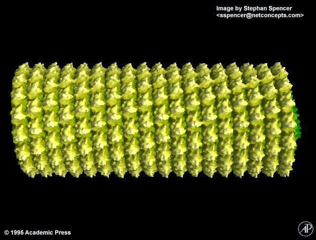 |
| TMV showing the helical arrangement of subunits |
 In contrast, the helical pitch of the subunits that form the outer casing of Tobacco Mosaic Virus illustrates that assembly pathways can influence the arrangement of subunits in a way that is distinct from the maize arrangement of its kernels. It is clear that evolution has settled (as usual) on several ways of packaging material either for protection (as in the viral capsid and nucleic acids) or for access (in the case of maize). Moreover, in contemplating assembly, the TMV arrangement has more in common with a certain Swedish domestic furnishing organisation, than a classical sculptor! Although the occurrence of natural helical growth is observed in some climbing plants (above right)?
 Those of you over 50, or interested in space exploration, will recall the LEM (Lunar Excursion Module) which was delivered to the moon in 1969 via Apollo 11. For many of us currently active in Science, this was surely a landmark event. The general features of the LEM are strikingly similar to those observed through many elegant Electron Microscopy studies of Bacteriophage T4, and more recently through the superb crystallographic work of Professor Michael Rossman's group at Purdue University. I think the comparisons speak for themselves!