Friday 31 January 2014

Guinea Pigs and fruit flies

What do guinea pigs, Saccharomyces cerevisiae, zebra fish and Escherischia coli have in common? They are all used experimentally to investigate Biological phenomena. We refer to them collectively as model organisms. So why Drosophila melanogaster and not Musca domestica? It usually comes down to practical reasons. Drosphila, or fruit flies have been used for scientific discovery for over 100 years. They are fast breeders, they eat cheap food and they are easy to store and maintain. From a genetics point of view, this fits the bill for mutational studies. Hence we have a rich history in using the fruit fly to explore the relationship between genotype and phenotype in general, but in particular phenomena such as eye colour and more recently development. In general, Biologists use the simplest organism they can, until a more complex one is required to investigate a phenomenon.


Horses for courses I. In my opinion, some of the greatest experiments in Molecular Biology have been carried out on bacteriophage and E.coli. The great ideas of Salvador Luria and Max Delbruck on evolution were a result of a statistical analysis of the frequency of mutations that led to bacteriophage resistance. So what does all that mean! Darwin's ideas suggested that mutations arise spontaneously in populations and it is only when those mutations give rise to some benefit to the organism, that they become fixed in a population, eventually out-competing the wild type organisms. In order to confirm this hypothesis, Luria and Delbruck turned to bacteria and bacteriophage (these are viruses that attack bacteria). Because the numbers of bacteria in a culture are in the millions, and the phage (as they are called) are even greater in number; and, doubling times are 20 minutes, it became possible to carry out a statistically controlled analysis. This demonstrated experimentally that Darwin's ideas were correct. This was only possible using this model organism.


Horses for courses II. Bacteria can provide us with insight into many molecular processes, but unlike human cells, bacteria have no nucleus (hence the name prokaryote). However, there         

are many aspects of human biology that would not be appropriate to try and explain from a study of bacteria alone. So, for example, if we want to understand the way in which humans develop, we turn to fruit flies, zebra fish and mice. The genes that are responsible for laying down the body plan for a fruit fly are essentially the same as those that dictate the positioning of our limbs and the polarity of our body (head and toes). These genes (including hox genes) are often called master regulators, since they coordinate sets of other genes and in particular their spatio-temporal patterns of expression (or simply when and where they are expressed). 

I have chosen to focus on the simple beetle Tenebrio molitor (or the mealworm), since it is easily obtained as a living larva, it is sold cheaply as dried food (for birds and humans), which simplifies the biochemistry; and it can be readily matured in the lab, producing a black beetle. Work in a number of genome labs have begun to explore the response of T. molitor to bacterial infection, and related genomes are available: the mealworm genome will be completed soon and we can already use its transcriptome to access information about its genetic makeup. Lets see if we can find out something new in our very own post-genomic study.


Wednesday 29 January 2014

The Importance of Evidence in Science

My school Chemistry text book told me that atoms are like the solar system: in which electrons orbited the central nucleus just like the planets in our own solar system orbit the sun. The truth is much more complex, and the recent announcement that the Higgs Boson (a particle that confers mass on other subatomic particles) has been "seen" using a high energy particle accelerator doesn't really fit with the simple model that I used to get me through my chemistry exams. But that's OK because it was an early stage in my journey to making discoveries of my own (or more accurately with colleagues) and I developed a level of understanding that enabled (or empowered) me to question established facts and concepts and to make up my own mind. This can only be achieved by looking at the "evidence" that has been used to justify a particular conclusion. So what is evidence and how do we evaluate it?

The Oxford English Dictionary defines "evidence" as follows:

                "The available body of facts or information indicating 
                 whether a belief or proposition is true or valid"

There is a problem with this definition, since a fact can be challenged by an experiment, which often happens as new technology emerges. So, until about 1985, we believed that all enzymes were proteins. Tom Cech and Sidney Altman, independently demonstrated that RNA, previously thought to be an information molecule, could also catalyse certain biological reactions. Not only has this subsequently been confirmed by many labs, but it has opened the way for a new way of thinking about genes and gene therapy. 


In this case the facts changed and therefore new evidence was used to displace the protein-centric idea. Facts in Science only remain set in stone after they are subjected to rigorous investigation. The double helix of DNA, now an iconic symbol, was thought to comprise 3 strands, until Watson and Crick (not forgetting of course Rosalind Franklin and Maurice Wilkins) demonstrated unequivocally, that it was a double helix...until of course we discovered Z DNA and quadruplexes at the tips of our chromosomes. But don't panic, double helical DNA is here to stay, it is just that when solid evidence is presented that challenges the universality of this structure it often explains things that were a little awkward to accommodate, in this case recombination and chromosome crossing over.


Geoff Schatz
So how do we deal with the "moving target"  or "shifting sand" that is the scientific evidence base? My first suggestion is to embrace it: always challenge a concept or fact by asking yourself this question. What is the definitive (or most compelling) experimental result that led to the formulation of a hypothesis? When Geoff Schatz was head of Biochemistry in the Biozentrum at Basel (Switzerland) he transplanted short segments of amino acids that he suggested were responsible for targeting proteins to the mitochondria (the organelle that generates cellular energy) onto cytoplasmic proteins. The result was that these proteins were now targeted to the mitochondria. This is referred to by Molecular Biologists as "an elegant" experiment: it cut to the heart of the problem and yet is very simple in concept. Of course Schatz was drawing on on years of experience, great insight, formidable technical skills and of course he was a bit of a genius! This concept remains robust, but there are exceptions: the important thing is that Schatz's (and others similar scientists) provided us with a model that allowed us to move forward in the lab in a logical and constructive manner. If the Schatz rules need to be overturned or modified by new and perhaps more compelling evidence, then so be it. This is how we progress in Science. Next, think about who judges the quality and significance of the evidence?


Monday 27 January 2014

The importance of planning experiments

Experimental Science. The journey from a student to becoming a practicing scientist never ends, or at least in my view it shouldn't. One of the things that I find most frustrating is a day spent with students carrying out an experiment while racing against the clock. Under these circumstances there is little chance of acquiring new skills (or even honing old ones) and an appreciation of the over-arching concepts, the significance of each step and ultimately an understanding of the outcome, are all lost in the process. In short the exercise is largely pointless. So, how should you approach your time in the lab? Below are some suggestions.

Listen, make notes, read instructions carefully and discuss your understanding with your group members. I generally find that taking notes during any meeting helps me remember things (and some say it aids in understanding). I recommend that you have a small notebook and pen with you when someone is giving a presentation in the lab or in a seminar (or in any meeting). If you have been given instructions or you are searching for information on the internet, read the instructions carefully. Avoid jumping into an experiment until you have mentally "walked through" all of the steps. 

Team-work. As a group you should then make sure you all understand what you have to do. Then allocate tasks and begin to get organised. Gather materials, clarify any uncertainties (after you have attempted to understand) with a demonstrator and write out a schedule for the day: we call these a work flow. This can form the basis of your methodology write up later.

Observation. I cant stress how important it is to keep watching what is happening to your samples, the amount of a solution that you dispense, the colour or appearance of a fraction from a column or a tube in an incubator. Again, make notes of what you observe. Some of these observations may not be important, but treat everything as important as you go through the experiment. It is only later that you can decide which observations should be included in the analysis of your results. 

Sample labeling. So far everyone has had an experience of losing their samples from one week to the next. Since we are running lab projects over several weeks, tracking your samples is vital. Think about how you label samples and don't leave it until the last minute. A petri dish labelled on the lid, is an experiment wasted if the lid is accidentally knocked off by you or a colleague in a shared fridge or incubator. Tubes labelled 1,2,3 or A,B,C will become forgotten among the multitude of As and Bs and Cs. If the sample is to be frozen, think about the likelihood of the label being lost. Use the right kind of pen/ink or sticker. Also write down the labels you have used and the location of your sample.

Tidy your bench. Before you leave, allow time to tidy your bench. This is considerate and polite, since someone else will have to do it for you: and that's unfair and unsustainable. Secondly, by tidying, you make sure that your solutions, samples and equipment are kept in the right place and temperature and that the equipment is well maintained. This is not an option.

Writing up your experiments. You have all been given clear guidelines for writing up your experiments. Take your time, think about the significance of what you have done and what you have observed. Experiments do not "work" or "fail", they give you outcomes or results that are influenced by many parameters: the reagent quality, the skill of your manipulations, proper use of instruments from pipettes to spectrophotometers etc. Therefore write up what happened and explain why you think you obtained your particular set of data. As you gain experience, your skills will improve and your ability to obtain robust data will also improve. This is a learning process and it never stops!

Tuesday 21 January 2014

Genomes and Bioinformatics at Y10

Today we have been using a method called BLAST searching. Very simply, we are accessing all of the genes and genomes whose sequences have been deposited in the "Public Domain". As young Life Scientists, this information will feature increasingly in your lives. As future professional scientists you will possibly contribute to our understanding of the significance of this information, in ways that we are unable to appreciate in 2014.

When I first helped sequence the gene encoding the restriction and modification enzyme from the lowly bacteriophage P1 in 1985, I had no idea that 30 years later, Mark Szczelkun at Bristol would use a new single molecule technique with this enzyme, to establish important new rules concerning the trafficking of molecules along the DNA double helix. Obtaining the sequences of genes and genomes was then a time-consuming labour of love. Recently, one of my own PhD students has recently obtained the sequences of 6 bacterial genomes using a University service in a matter of weeks: obtaining the sequence of two genes took 6 of us over one year! We are hoping to find a clue to the origins of a new genetic damage phenomenon which is linked to some cancers: chromothripsis. When we have analysed the data, you will be the first to know!


Returning to genomics and bioinformatics; I explained that the sequences of the genomes of man, mouse and pig were very similar (but each of their brains are quite different!). Interestingly, if Charles Darwin had used BLAST searching, I wonder if he would have developed his unifying theory of evolution. I find it easier to tell a chinchilla from a pig by looking at them, than I would by searching the data base. It is  much harder
to spot the difference pair of genes from mouse and pig and man than you might think. It seems that small differences in individual genes combine to determine the appearance, or "phenotype" of the organism (and indeed each cell). In higher organism it is a combination of the genome and our neural networks, that confers our evolutionary advantage.


The freedom to access to this phenomenal resource of biological data is, in my view, one of the greatest achievements of Science in this century. The challenges that lie ahead to interpret these sets of data will form a major part of your lives in Science over the next 50 years. I will leave you by asking you to think about evolution in terms of the complexity of the human genome and the success of the relatively simple genomes that are required to make viruses. Is the virus the most highly evolved? I hope you find time to enjoy searching through the NCBI portal.

Monday 20 January 2014

Oranges and lemons and quantum biology in Y12

The Rosy Periwinkle
from Madagascar
In yesterday's session, you set out to develop a series of plant extracts in order to test their anti-bacterial properties. This is a form of "Bioprospecting", a generic term sometimes used to describe the traditional approach to Drug Discovery. So for example, anti-cancer drugs such as vincristine and vinblastine have been found in the Rosy Periwinkle (left) found in Madagascar. Often these initial observations are based on local knowledge and cultural practices. Pharmaceutical companies have historically investigated such compounds and then developed methods for their synthesis in order to control the costs of production and to ensure the stability of supply. There are of course important issues relating to the impact on the communities who might be adversely affected by such discoveries. Indeed morphine, which can be found in poppy seeds is on the one hand invaluable in pain management, but on the other hand a major cause for social unrest in view of its illegal use as a narcotic, which in a similar way to cocaine impacts on certain developing countries such as Afghanistan.

Whilst I was homogenising the lemons, I was immediately transported to my
organic chemistry classes as an undergraduate at Sheffield and I remembered the story of oranges and lemons and the chirality of limonene. The characteristic difference between the smell of oranges and lemons couldn't be more subtle, from a chemical perspective. As you can see left the two molecules differ only with respect to their chirality. That is, both molecules are mirror images, like left and right hands. But that's clearly enough to change the way our brain interprets their smell, or odour. Which brings me nicely on to "quantum biology". 

It has been suggested that the translation of the information from a small molecule such as limonene into a sensory perception is partly a result of the shape of the molecule, but is possibly a result of the molecule's quantum level vibrational fingerprint! So as you know, all bonds have a characteristic set of vibration frequencies derived from bending, stretching etc. You may have come across this in Chemistry. These phenomena can be understood using quantum theory and unlike the lock and key concept (which we will explore soon) it has been suggested that neuronal responses to odour are triggered by quantum vibrations. So why would a good experiment be to compare the sell of limonene in both chiral forms with a deuterated version?

Sunday 12 January 2014

Pigments of my imagination

Some of the most enjoyable experiments last term involved the use of coloured solutions ranging from copper sulphate, for demonstrating the relationship between light absorbance and concentration to column chromatography. We also used milk to demonstrate the way in which salts such as ammonium sulphate and acids, such as acetic acid, could be used to fractionate proteins. Of course we also used ion exchange chromatography to separate proteins in milk on the basis of their surface charge. And of course, as you know milk is white. 


So why are some metal salts colourless in aqueous solution and why are dyes (and the salts of some metals, such as copper sulphate) red, orange, green etc? Recall that we selected a specific wavelength of light to measure the relationship between the colour intensity of copper sulphate, this is because the specific arrangement of electrons in these compounds (or molecules) leads to the absorbance of certain wavelengths of light and the emitted light is now coloured. This relationship between light (photons) and matter is at the heart of biology in the form of photosynthesis. Refer to you class sheets for the structures of the dyes that we used: you will notice that they all contain aromatic rings and these ring structures contain "delocalised" electrons which often (but not always) give rise to the spectral characteristics of the molecule. Since the energy of a photon is inversely proportional to its wavelength. light of shorter wavelength has more energy than longer wavelengths and we can draw conclusions about the energy states of molecules from such experiments. This is again a major aspect of the Biophysics of photosynthesis. 

The importance of many vitamins in Biology is a result of their role at the heart of many proteins including haemoglobin (which contains a form of porphyrin and iron, called haem) which transports oxygen around our cardiovascular system. Some enzymes are bright yellow in colour, owing to the presence of a flavin molecule, which facilitates the chemistry of catalysis that sometimes cannot be carried out by amino acids that make up the active site of the enzyme. You will also come to realise that energy in the form of the molecule ATP (adenosine triphosphate) is synthesised in the mitochondria following our metabolism of carbohydrates and fats via a series of complex events driven by electron flow between the haem components of a group of proteins called cytochromes. This process, which was first properly understood by the British
Biochemist Peter Mitchell, from his private laboratory on Bodmin Moor and around 30 years later, another British Biochemist, John Walker provided an important answer to the molecular framework of this phenomenon, when he determined the structure of one of the first molecular motors that couples electron and proton flow to ATP synthesis: the mitochondrial ATPase is shown on the right (or below). Isn't it an amazing molecule! Both scientists shared in separate Nobel Prizes. So understanding dyes can help us appreciate the way in which energy in the form of electrons within molecules can help us appreciate how Nature is able to mobilise energy from food and sunlight.


Friday 10 January 2014

Bleach: past fortunes and modern molecular biology

Thinking about the results of your experiments on the inhibition of bacterial growth by antibiotics, ampicillin and chloramphenicol, led me to look a little further into the chemistry and biology of bleach. What I hadn't realized is that bleach manufacture is something we do ourselves to protect against microbial attack! So the industries that grew up around Merseyside at the dawn of the industrial revolution (a life size model of a bleach packer is shown on the right, from the Catalyst Museum at Widnes) have been going on inside our cells for a long time before then. This is one of those "dangerous" processes that provides a defence for the body, but is also potentially hazardous. Just as the bleach packers from 150 years ago "held their lives in their hands", so do we when we selectively release activated chlorine via hypochlorous acid production from blood neutrophils to combat infection.


So what is the chemistry of bleaching? Y12 chemistry classes this week will cover the reaction through which sodium hypochlorite (usually in conjunction with the alkali sodium hydroxide) generates chlorine and atomic oxygen, both of which have anti-microbial properties. The effects are not however, non specific (like corrosive compounds such as strong acids and alkalis): it appears that bacterial proteins become unfolded in the presence of the hypochlorous acid and this activates a class of protein called molecular chaperones (left). These proteins were originally identified as heat shock proteins: they are switched on in response to environmental stress. They protect our key enzymes and other proteins for a short while until the danger has passed. And of course the danger does pass when the agent is a gas. So when the bleaching effect of chlorine subsides some microbes begin growing again. This is whey we see growth in the cultures to which bleach was added, but not in the cultures where the antibiotics were added. We really need to do a systematic study of the concentrations of bleaching agent, the number of cells in the inoculum and the growth rates. Let's do this over the next couple of weeks


Since this discovery, it has become clear that Neutrophils (cells that form part of our immune system) produce hypochlorous acid from chloride ions (found everywhere in the body: think of salt) using the enzyme myeloperoxidase and hydrogen peroxide (another popular hair bleaching agent that parents may know!). The enzyme has a molecule of haem at its centre, similar to that found in haemoglobin, so it is a coloured protein, but unlike haemoglobin, it is green. In fact the green colour of pus is due to secreted myeloperoxidase!


What next in the bleach story? Well Ursula Jakob (left) at the University of Michigan has discovered a gene sensor called the Reactive chlorine receptor, that switches on the genes that encode the molecular chaperones above. This means that bacteria defend themselves against bleach until they become overwhelmed. So this is another example of a "biological arms race" that we find in Nature and is a great example of evolutionary adaptation. However I think this observation suggests that bleach resistance could emerge and this would cause more problems for human health! Little did the bleach manufacturers of old realise when they set out on the road to develop disinfectants and solutions for the textile industry.  I think we should take a further look at antimicrobials such as bleach in more detail this term! Any thoughts?

Thursday 9 January 2014

Microbes in sickness and health, antibiotics and the importance of accurate pipetting!

In the Y10 class today, we began the first week of a project aimed at exploring the growth of microbes and the experimental methods for exploring the properties of antibiotics and disinfectants in eliminating bacterial and fungal contamination. The first experiment was straight forward: how do the antibiotics ampicillin and chloramphenicol compare with detergent and bleach in slowing (or killing) a vigorous culture of E.coli? What became clear at the end of last term, was the challenge of accurately pipetting microlitre volumes and was in evidence today, was a similar story. We need to master our pipetting techniques and take greater care in handling samples!

There are a few rules when dealing with small volumes of reagents and sterile tubes containing culture media and bugs. Never tip the tube upside down, if you need to mix, use the vortex! Never tip an eppendorf tube upside down when it contains only 20ul of sample: if you do, pulse it in the microfuge to get the contents back to the bottom of the tube. You will not be able to do reproducible molecular biology if you keep repeating these basic errors. When we did the pipetting test in the middle of the day, over 60% did not obtain perfect results. Let's get on top of this!


Getting back to microbes, I hope you all saw the Penicillium I had grown over the holiday. You have inoculated broths at room and fridge temperatures. They are coming on nicely and we will work with them next week. The penicillin story is another one of those great stories of accidental, or serendipitous, discoveries. That is you stumble across an unexpected observation and then pursue its significance. There is no doubt that Fleming's discovery has had a major impact on world health. I hope you enjoy completing the tasks.

I just spent an hour looking over the growth results from Tuesday and everyone has obtained the same, interesting result. However I will not give away the "score" until you have all looked. So until next week....

Tuesday 7 January 2014

Genomes and their value in Biotechnology

During the first part of the term all Y12 students were allocated a bacterial genome, following a number of lab sessions on how to use BLAST (Basic Local Alignment Search Tool) through the NCBI (National Center for Biotechnology Information). We first looked at the logic by which the BLAST algorithm compares a string of amino acids (in the form of the one-letter amino acid code) from one genome with another (BLASTP) and also at the DNA coding level (in the form of GAT or C). Remember that the searchable data sets are dynamic and that the computational methodology draws on a knowledge of protein chemistry and comparative sequence studies: it isn't quite like searching for a phone number: BLAST gives you not only a precise match, but also similar sequences (this isn't the case if you dial the wrong mobile number!)


In both cases, this powerful method rapidly enables a research worker to compare genes from different organisms and answer questions relating to biology (the evolutionary relationships between individual genes or whole genomes) and chemistry (the level of tolerance between the sequences of an enzyme or a gene regulatory molecule to mutation can give clues to the functional parts of a molecule). These are just two examples of how genomic data can help a research scientist. Indeed, it has been suggested by some influential scientists and commentators that experiments at the bench will become less common, as we learn to interpret genomic (and large data sets relating to proteomes and transcriptomes which are often called "omics"). 

I am not sure that Biology will become an in silico pursuit just yet, but it is certainly the case that as we accumulate knowledge of the components of cells and tissues and their interaction networks, the areas of Systems Biology and Synthetic Biology will become increasingly powerful and predictive. Wouldn't it be nice to be able to use Bioinformatics to design a new molecule or anew organism without spending time and money carrying out preliminary experiments? 


You should look at your allocated genome and ask the following questions.

Why was it sequenced? (Does it have a basis in medicine or some commercial interest beyond the basic Science value?) Are there any phenotypic features that make it interesting to compare with other organisms? Does it have any unique genes (or genes shared by a niche group)? Does it exhibit any epigenetic phenomena? 

Choose any pathway such as a metabolic pathway (the Krebs Cycle for example), or a regulatory pathway (gene activation or repression, for example the Trp operon), a biosynthetic process, such as protein synthesis, RNA synthesis, DNA synthesis, cell division or cell growth and map out the genes and proteins involved. Then compare these genes selectively with the rest of teh published genome data and try and place your organism into an evolutionary and functional context. If you want to make a start why not look at DNA replication, since all organisms have to do it and we looked at the DNA polymerases during the class. Recall, there are often several DNA polymerases found in prokaryotes, but some are useful but not essential. How similar are replication systems in prokaryotes and eukaryotes? In other word what difference if any doesa a nucleus make?


Sunday 5 January 2014

The coming term...

Penicillium mold
Now that you are all familiar with the general layout of the Innovation labs and have got used to the use of the lab note book system, I have been working with the teaching staff to produce a system that will allow you to build on the skills developed in the first term and take on some more challenging Science projects.  The three labs will be overseen by a member of the Science staff who will begin the formal monitoring of your  lab note books: we are aiming to move from your induction term to a fully professional research lab environment. I am delighted to say that this is a result of the responsible and committed way that you all conducted yourselves in Term 1, as well as the speed with which you mastered some rather complex methods and ideas. You have made spectacular progress, so I am going to challenge you even more!

Cyanobacteria
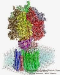
This term, Y12 students will extract and fractionate a range of compounds from natural sources and investigate their anti-bacterial and anti-fungal activities compared with commercial compounds. This will be followed by an ambitious project in which we shall adopt the concepts of "Synthetic Biology" to engineer novel, recombinant peptide synthesising enzymes from cyanobacteria. For this project we will need to draw on your Bioinformatics skills from Term 1, together with the methods of recombinant protein production from the Eden project. We will also be firing up the PCR machines in order to generate the appropriate coding sequences from cyanobacterial genomic DNA. Now that Dr. Moore, has recovered from the shock, our new lab layout should help us meet the challenges of this exciting new project. 

The Y12 projects form part of our relationship with our regional partners: 

Unilever: novel therapeutics from natural products.


Unilever produce more than 400 brands focused on health and well-being: their portfolio ranges from nutritionally balanced foods to ice creams, soaps, shampoos and everyday household care products. Some of teh brands you may know are: Lipton, Knorr, Dove, Hellmann's and Omo.


Croda:  Synthetic Biology.

Croda International is a global leader in speciality chemicals, sold to a wide range of markets- from Personal Care to Health Care; from Crop Care to Coatings and Polymers.


The Y10 students will build on the introductory microbiology work from last term by investigating the way in which microbes can be controlled using general agents such as bleach and the more sophisticated control used for therapeutic purposes: antibiotics such as penicillin, kanamycin, tetracycline and chloramphenicol.  We will be supported in this work by ProLabs who were so generous in supporting and resourcing our microbiology lab classes last year.

Using broth and plate culture techniques, we will investigate the tolerance of bacteria to mainstream antibiotics and look at the properties of commercial cleaning agents with respect to different classes of organisms. We shall use a combination of laboratory strains and we shall also repeat the Fleming experiment, by isolating Penicillium mold and using the organism and extracts from it, to reproduce Fleming's early experiments on antibiotics.

We shall begin with an investigation of the concept of dosage. How much anti-bacterial agent does it take to kill a culture of live bacteria? Is it sufficient to kill 99.9% of germs, and what are the pros and cons of administering antibiotics? These issues will be explored alongside the basic chemistry underlying the active ingredient(s) and mode of action of commonly used antibacterial agents.


Saturday 4 January 2014

Blood, sweat, tears and the milk of human kindness! Part 2

So what's in the title? Well, we started the year looking a the differential growth of oral swabs on blood and nutrient agar plates (and blood forms the basis of Y11's innovation work in term 1 and part of the introduction to forensics for Y12s after the Easter vacation). Sweat and tears? Well, we use lysozyme to break down the bacterial cell wall (and you will recall that's where we secrete lysozyme, as a primitive anti-bacterial defence mechanism). And that leaves milk, which needs no further explanation!

So moving on to the Y12 project. Early discussions with senior members of the Eden Biodesign team, significantly enhanced the design of the Y12 project that centred on the controlled production and purification of a recombinant protein in a bacterial host. Eden (for short) produce recombinant polypeptides (another name for proteins) at level of purity far exceeding that achieved in our labs (since their products are often administered to patients!). We decided however, that the best initial approach was to introduce you to the fundamental principles of cell growth, gene regulation, selective gene induction, protein isolation and purification in a simple prokaryotic system first! Well, I can assure you that all of your final presentations and those selected for presentation to the Eden team, were really impressive. 

In one sentence I have covered a significant part of an undergraduate degree course in Molecular Biology (an observation that was not unnoticed by the Eden team). So let's just take a few lines to explain the Science behind this project.

Escherichia coli were used as the bacterial host for the expression of a recombinant form of the Green Fluorescent protein, which is naturally expressed in certain jelly fish. One of my colleagues at the University of Sheffield, Professor Simon Foster passed on a bacterial strain harbouring a plasmid encoding this gene which is placed downstream of the lac operon. When you established that the optimum growth temperature was 37 degrees for the strain, you then induced expression of the GFP gene by "de-repressing" the lac promoter. Recall that the IPTG (a molecular mimic of the sugar lactose) prevents the lac repressor from blocking access to the host cell RNA Polymerase. As a consequence the gene for GFP is expressed, the protein is synthesised from its mRNA (on the ribosome) and by the end of week two, you all had a batch of cells in the freezer packed full of recombinant GFP.

A range of NTA columns containing
different immobilised metal ions
So far so good. Time to move from Molecular Microbiology to Biochemistry (and since its my blog, the most important Life Science subject!). One of the tricks that have made Biochemistry so accessible (in particular protein biochemistry) to general molecular biologists, has been the application of genetics to add "tags", or additional sequences of amino acids that selectively bind to "ligands" (defined molecules ranging from metabolites to other proteins) that can be readily immobilised (attached) to the beads that make up column chromatography resins. We call this methodology affinity chromatography and we used the interaction between a poly Histidine tag (6 His codons were added to the GFP gene) and Nickel ions immobilised through a compound called NTA. This interaction is the basis of one of the most popular methods in protein purification in Biotechnology today. 

So, we have a cell pellet in the freezer, stuffed with GFP (His-tagged): the next step is to release the GFP (along with E.coli's own proteins that can be anything up to 2-3000 in number!). To do this we chose to add the enzyme lysozyme (obtained from egg white). It is possible to break cells by a range of chemical and mechanical methods, and you will use different methods in the Unilever project in January. Lysozyme is also an important landmark enzyme in the history of Biochemistry, since it was the first enzyme to have its 3D structure determined (above) by Sir David Phillips' group at Oxford in 1965. 

The next step in the project was to use a column of Ni-NTA beads to capture the soluble recombinant GFP, while removing the contaminating host proteins. This was probably the point at which some of you went astray (although I estimate from the SDS PAGE experiment that more than 70% of the class obtained pure GFP). The difference was mainly the yield; which is commonly the case if you ask a new PhD student to make a batch of the lab's favourite recombinant protein, so nothing to worry about for a first attempt!

Several of the class preparations
of Ni-NTA purified GFP.
(Lane 6 in the middle is a marker)
The tricky part of column chromatography is ensuring the resin stays fully immersed in buffer and that the sample loaded is not too dilute. In both ion exchange and affinity chromatography (unlike gel filtration chromatography), the material being purified is adsorbed to the resin and therefore becomes concentrated. This means you can get away with less attention to detail than when performing gel filtration chromatography, where the sample becomes increasingly dilute as it runs through the column. The elution step involved the displacement of the GFP bound to the Ni ions via the hexa-His tag. For this we used a high concentration (300mM) of imidazole (a mimic of His). The GFP was selectively displaced and samples collected in eppendorf tubes in 2-3 drop fractions. Again, this is challenging and I would say that around 10% of you obtained the GFP in a form that was sufficiently concentrated to see it glow in the lab. However as in the SDS PAGE gel at the left, most of the class produced an almost pure sample and I (and the Eden team) were duly impressed.

After you had all left Michael pooled the samples and we used a larger column to concentrate the "class GFP". As you can see below, one of my best Christmas presents was the bright, green preparation that was a fantastic achievement by you all!

A "class" act!