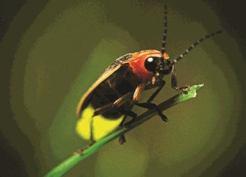
 The summer brings out the best in most of us (purely based on the evidence of the greater number of smiling people on the platform when I catch the train!). So, I thought about choosing a molecule that reflected this mood. I could have looked amongst the proteins that are the targets of psychotic drugs, or I could have gone for sunlight capturing molecules involved in photosynthesis. In January of 2016, I discussed a number of photo-activated proteins after a thrilling seminar from the Biochemist Tomas Carrel. See here. This month I have chosen the enzyme luciferase, a key element in the generation of light in insects such as the glow worm and the fire fly (Photinus pylaris). I hope you agree that these creatures (notwithstanding the general unpopularity of most insects), make most people smile!
luciferin + ATP → luciferyl adenylate + PPi
luciferyl adenylate + O2 → oxyluciferin + AMP + light
Light is produced because the reaction forms oxyluciferin in an electronically excited state. The reaction releases a photon of light as oxyluciferin returns to the ground state (in this case, the "quantum mechanical" state of a system having the lowest possible potential energy. The expression is also used in Biochemistry to define the lowest free energy state of substrate(s) in an enzyme catalysed reaction, usually with respect to the transition state and the products of the reaction). Firefly luciferase generates light from luciferin in a multistep process. First, D-luciferin is adenylated by ATP to form luciferyl adenylate and pyrophosphate. Following this "activation" by ATP, luciferyl adenylate is oxidized by molecular oxygen to form a dioxetanone ring. A decarboxylation reaction yields the excited state of oxyluciferin, which tautomerizes between the keto-enol form (at a given pH and temperature, all carbonyls have a tendency to shift between these two forms: you can read more here). The reaction finally emits light as oxyluciferin returns to the ground state. [I shall return to the important topic of "excitation" of molecules and its importance in Biological systems in a separate post.]
This is quite a complicated phenomenon without a background in undergraduate Biochemistry, Chemistry or Biophysics, so don't worry if it leaves you a little baffled. Think of the dyes that colour your clothes, or a bright blue copper sulphate solution. It is sometimes possible to re-organise electrons in a molecule in response to visible and uv light. This can result in a portion of the visible spectrum being removed by the molecule, which results in a very specific colour of a solution of the molecule. The process involves light energy in the form of photons, re-organising specific electrons in the molecule, followed by their return to the "unexcited" state which can be accompanied by the emission of a colour change, a fluorescence emission or phosphorescence. There are specific "pathways" that are described in quantum mechanics that account for these phenomena and why different molecules choose one over the other, or none at all! I shall attempt to write a post on these important phenomena in the near future, since they are particularly important in the mechanism of photosynthesis.
This is quite a complicated phenomenon without a background in undergraduate Biochemistry, Chemistry or Biophysics, so don't worry if it leaves you a little baffled. Think of the dyes that colour your clothes, or a bright blue copper sulphate solution. It is sometimes possible to re-organise electrons in a molecule in response to visible and uv light. This can result in a portion of the visible spectrum being removed by the molecule, which results in a very specific colour of a solution of the molecule. The process involves light energy in the form of photons, re-organising specific electrons in the molecule, followed by their return to the "unexcited" state which can be accompanied by the emission of a colour change, a fluorescence emission or phosphorescence. There are specific "pathways" that are described in quantum mechanics that account for these phenomena and why different molecules choose one over the other, or none at all! I shall attempt to write a post on these important phenomena in the near future, since they are particularly important in the mechanism of photosynthesis.
The protein molecule (shown left from a dinoflagellate) comprises two major structural units. The blue (mainly) beta barrel sits beneath the alpha-helical arrangement, with the adenylate and the chromophore positioned at the junction of the two domains. On binding the reactants the domains come together to exclude water, which increases the half life of the "excited" state of the oxyluciferin. The details vary a little from species to species and this leads to a variation in the wavelength of the emitted light. One mechanism proposes that the colour of the emitted light depends on whether the product is in the keto or enol form. The mechanism suggests that red light is emitted from the keto form of oxyluciferin, while green light is emitted from the enol form of oxyluciferin. This is not proven, but the logic relates to the well established connection between resonance structures and the energetics of absorption of light in the visible and uv spectrum. There are some other ideas, but even though a consensus hasn't yet been reached, all mechanisms will probably connect the local (molecular) environment with the stabilisation of the excited state (see below RHS).
 You may wish to compare the properties of luciferases with naturally fluorescent proteins such as the Green Fluorescent Protein (GFP for short). Can you think of the biological advantages for an organism emitting light? Maybe a useful exercise is to compare and contrast the applications of these enzymes in contemporary experimental molecular cell biology? Can you find glow worms and fireflies in the UK? Take a look at the survey.
Key points from the Blog. There are naturally occurring proteins (and small molecules) that have fascinating optical properties. Such properties are sometimes related to their requirement for energy to drive unfavourable reactions (DNA repair, photosynthetic electron transfer). In some cases, the natural glow of a fire-fly or the bright fluorescence of marine organisms has evolved for reasons that are not entirely understood. However, such beautiful natural phenomena attract Biochemists and they can lead to technologies that unlock hidden secrets in the behaviour of cells. Luciferases are used in a wide range of diagnostic and research methods, and I hope you agree with me that they are incredible molecules, however I think GFP currently holds the prize as the most important optical probe in contemporary biology.


