 The
Brain, was first observed directly (in a properly documented way) in
humans around 500 years ago, after it became acceptable (again) to
desecrate the human form, in order to try and obtain some insight into various diseases, in particular the Black Death. Leonardo da Vinci (the famous Renaissance polymath) is often associated with the first work, but Al-Hazan, a scholar from Basra in modern day Iraq, made important experimental observations, especially in respect of vision, 500 years earlier. The ancient Greeks carried out the first documented dissections,but this practise became taboo with the spread of Christianity and Islam. The re-emergence of dissection of cadavers (not an intuitive word for a corpse, probably derived from the Latin verb cadere, to fall: think of "the fallen" used to describe the victims of a battle) is largely associated with Viselius and Leonardo at the time of the Renaissance. There were certain characteristics needed to become an anatomical recorder, in the 16th Century. A strong stomach, a strong arm, a keen eye for detail and a brilliance at drawing. If you haven't seen any of Viselius's or Leonardo's anatomical drawings, search Google images without further ado! Both Viselius and Leonardo captured the underlying elements from which the human body is sculpted and shaped with great skill: all artists from portrait painters to cartoon animators study these pioneering works to this day.
The
Brain, was first observed directly (in a properly documented way) in
humans around 500 years ago, after it became acceptable (again) to
desecrate the human form, in order to try and obtain some insight into various diseases, in particular the Black Death. Leonardo da Vinci (the famous Renaissance polymath) is often associated with the first work, but Al-Hazan, a scholar from Basra in modern day Iraq, made important experimental observations, especially in respect of vision, 500 years earlier. The ancient Greeks carried out the first documented dissections,but this practise became taboo with the spread of Christianity and Islam. The re-emergence of dissection of cadavers (not an intuitive word for a corpse, probably derived from the Latin verb cadere, to fall: think of "the fallen" used to describe the victims of a battle) is largely associated with Viselius and Leonardo at the time of the Renaissance. There were certain characteristics needed to become an anatomical recorder, in the 16th Century. A strong stomach, a strong arm, a keen eye for detail and a brilliance at drawing. If you haven't seen any of Viselius's or Leonardo's anatomical drawings, search Google images without further ado! Both Viselius and Leonardo captured the underlying elements from which the human body is sculpted and shaped with great skill: all artists from portrait painters to cartoon animators study these pioneering works to this day.I liked Leonardo's attempt to work out what, if anything surrounded the brain inside the skull, gave rise to its characteristic "human" properties, including thoughts and memories. He made two precise incisions one from which he would collect any fluid (from a corpse, I should add!) and the other into which he injected molten wax. As the hot wax seeped into the cavity, out came any internal fluid. As soon as wax began to appear at the outlet hole, he would stop. The skull was then carefully removed and the wax cast provided a replica of the space between the brain and the inner surface of the skull. Needless to say little was gained from this experiment, which just goes to show that even geniuses have a bad day at work!
 The detailed drawings of the brain, resulting from the first observations of its anatomical features, immediately stimulated ideas about what we now refer to as structure-function relationships (or sometimes called structure-activity relationships, particularly in the field of Chemistry). The organs of the body are all rather different. The liver,
the heart, the eye, the ear etc, are all the products of evolution and
have become specialised "machines" for handling our food and waste (the liver, not forgetting the kidneys), blood circulation (the heart), vision and hearing (the eyes and ears respectively). Maybe I should think about an "organic" blog? Back to the brain for now. As you can see from the demonstration by a contemporary artist (above), the brain is roughly oval shaped (in three dimensions this approximates to an oblate spheroid), with a stem, that connects the brain to our circulation (blood supply), but most importantly the nervous tissue emerging from the stem travels down the spinal chord to "innervate" the body. So the first structure function observations are that the brain is on the top of our heads, fed by a blood supply pumped around by our heart and its influence on our movement and mood is the result of a cable that is distributed throughout the body's organs and tissues. You will have heard the expression "nerve centre" used to describe the most important part of an organisation, for example, the Cabinet Office at Number 10 Downing Street is said to be the "nerve centre" of British government, or the "nerve centre" of any airport is the "traffic control" tower. We also use the expression, "at the heart of an organisation", similarly this organ feeds the body by pumping nutrients around and removing waste products. The brain sends signals and issues commands, while the heart keeps you in good order. You can begin to see perhaps how scientific terms have crept into our everyday language, and usually for good reason. [Can you think of any others?]
The detailed drawings of the brain, resulting from the first observations of its anatomical features, immediately stimulated ideas about what we now refer to as structure-function relationships (or sometimes called structure-activity relationships, particularly in the field of Chemistry). The organs of the body are all rather different. The liver,
the heart, the eye, the ear etc, are all the products of evolution and
have become specialised "machines" for handling our food and waste (the liver, not forgetting the kidneys), blood circulation (the heart), vision and hearing (the eyes and ears respectively). Maybe I should think about an "organic" blog? Back to the brain for now. As you can see from the demonstration by a contemporary artist (above), the brain is roughly oval shaped (in three dimensions this approximates to an oblate spheroid), with a stem, that connects the brain to our circulation (blood supply), but most importantly the nervous tissue emerging from the stem travels down the spinal chord to "innervate" the body. So the first structure function observations are that the brain is on the top of our heads, fed by a blood supply pumped around by our heart and its influence on our movement and mood is the result of a cable that is distributed throughout the body's organs and tissues. You will have heard the expression "nerve centre" used to describe the most important part of an organisation, for example, the Cabinet Office at Number 10 Downing Street is said to be the "nerve centre" of British government, or the "nerve centre" of any airport is the "traffic control" tower. We also use the expression, "at the heart of an organisation", similarly this organ feeds the body by pumping nutrients around and removing waste products. The brain sends signals and issues commands, while the heart keeps you in good order. You can begin to see perhaps how scientific terms have crept into our everyday language, and usually for good reason. [Can you think of any others?]  The next things that you notice, if you pick up a human brain, is its weight, its colour, its texture and its surface nooks and crannies (gyri and sulci), all of which which are often likened to pasta, in particular spaghetti. In fact the contours of the surface tissue (the gyri and sulci, were originally thought to be responsible for higher and lower intellectual powers by
early anatomists). If you now turn the brain over, you will see the
business end that connects the body of the brain to the rest of the
body, via the brain stem. Just think of that scene the movie "The Matrix" when our computer whizz is physically connected to "the Matrix" by Morpheus. The same logic applies. Finally, I should add that the brain is surrounded by a membrane, which is called the meningeal membrane, or just the meninges (meninx is ancient Greek for a membrane and the meninges actually comprise three layers of membrane, as shown above LHS). It is this set of membranes that becomes inflamed when either a virus (such as Herpes simplex or Varicella zoster) or a bacterium (including, Neisseria meningitidis, Streptococcus pneumoniae and E.coli) invades and gives rise to the very serious disease you will all know as meningitis (literally inflamed membranes around the brain).
The next things that you notice, if you pick up a human brain, is its weight, its colour, its texture and its surface nooks and crannies (gyri and sulci), all of which which are often likened to pasta, in particular spaghetti. In fact the contours of the surface tissue (the gyri and sulci, were originally thought to be responsible for higher and lower intellectual powers by
early anatomists). If you now turn the brain over, you will see the
business end that connects the body of the brain to the rest of the
body, via the brain stem. Just think of that scene the movie "The Matrix" when our computer whizz is physically connected to "the Matrix" by Morpheus. The same logic applies. Finally, I should add that the brain is surrounded by a membrane, which is called the meningeal membrane, or just the meninges (meninx is ancient Greek for a membrane and the meninges actually comprise three layers of membrane, as shown above LHS). It is this set of membranes that becomes inflamed when either a virus (such as Herpes simplex or Varicella zoster) or a bacterium (including, Neisseria meningitidis, Streptococcus pneumoniae and E.coli) invades and gives rise to the very serious disease you will all know as meningitis (literally inflamed membranes around the brain).  Now we have to move from the first phase of observation, to looking inside the brain, and of course this involves
dissection or increasingly imaging. But it was necessary to dissect the
brain first in order to enable us to interpret the so called brain scans that we read about today and which some of you may have experienced (more on brain scanning later). The field of anatomy is critical in medicine,
if you are carrying out surgery, you need to know where to make a hole
(or initial incision) to get to the heart, the kidney, the lungs, but
the brain is actually pretty straight forward. Then as a young doctor, you have to know the names of all the parts: you may be working as a team and you need to specify where to cut or where to stem blood flow etc, everything in medicine has a name. Unfortunately, the name is not always easy to understand. Since most of the body parts were given names in Latin, they are not obvious to English or Japanese speakers! The same is true of a car. The parts of the engine don't have everyday names: the carburetor, the piston, the spark plug, the distributor, the clutch etc. These words sound less foreign to us perhaps, but they need memorising if you want to be a mechanic. But now I make the case for the vet, who has to learn the anatomy,
not only of humans (although s/he doesn't usually have to treat
humans!) but of the ostrich, the cow, the dog, the lizard etc. For my
money vets are rather interesting individuals and impress me enormously with their capacity to retain information in a spatially defined way. Hence the demanding A level entry requirements!
Now we have to move from the first phase of observation, to looking inside the brain, and of course this involves
dissection or increasingly imaging. But it was necessary to dissect the
brain first in order to enable us to interpret the so called brain scans that we read about today and which some of you may have experienced (more on brain scanning later). The field of anatomy is critical in medicine,
if you are carrying out surgery, you need to know where to make a hole
(or initial incision) to get to the heart, the kidney, the lungs, but
the brain is actually pretty straight forward. Then as a young doctor, you have to know the names of all the parts: you may be working as a team and you need to specify where to cut or where to stem blood flow etc, everything in medicine has a name. Unfortunately, the name is not always easy to understand. Since most of the body parts were given names in Latin, they are not obvious to English or Japanese speakers! The same is true of a car. The parts of the engine don't have everyday names: the carburetor, the piston, the spark plug, the distributor, the clutch etc. These words sound less foreign to us perhaps, but they need memorising if you want to be a mechanic. But now I make the case for the vet, who has to learn the anatomy,
not only of humans (although s/he doesn't usually have to treat
humans!) but of the ostrich, the cow, the dog, the lizard etc. For my
money vets are rather interesting individuals and impress me enormously with their capacity to retain information in a spatially defined way. Hence the demanding A level entry requirements! So back to brain dissection. By making clearly defined incisions and slices, early anatomists elucidated the basic structure of the brain and identified the main features of the blood supply and output cables, the nerves. With the introduction of high magnification lenses and microscopes, our understanding of structure and function in the brain emerged rapidly throughout the Renaissance and up to the 19th Century, when a major shift in experimental Biology took place and it was from the field of Physics, rather than Biology. The basic layout of the circulatory system was worked out by pioneers like William Harvey, but how did the brain actually work? Some important clues came from the emerging field of electricity, and from a rather beautiful part of the world, Italy, where a couple of "obsessively competitive" Italian scientists were trying to understand the connection between electricity and living organisms. Professor of Anatomy at the University of Bologna (the most ancient of European Universities) Luigi Galvani was making waves with his observations of frog muscle contraction in response to electrical stimulation. Curious to understand these dramatic experiments, Alessandro Giuseppe Antonio Anastasio Volta, from northern Italy, where the sublime Lake Como creates a border with Switzerland, had risen to the elevated position of Professor of Physics at the University of Pavia, by 1779. Volta, was having a few issues with Galvani's interpretation of his observations. A scientific stand-off ensued, but the outcome was a good one. It was finally agreed that muscle tissue could conduct electricity and that the signals that would normally be generated in the brain to induce muscle contraction, were electrical. Volta's contribution was to clarify the fact that the development of a potential difference (now measured in volts, after Volta), was simply a Biological version of what was understood from chemistry. This all paved the way for our understanding 100 years later of the mechanism of action of nerves: specialised cells that combine electrical conduction
with chemical transmission. I wont go into it here but the work of
Sherrington and Lord Adrian were key to understanding these processes
and are discussed here.
So back to brain dissection. By making clearly defined incisions and slices, early anatomists elucidated the basic structure of the brain and identified the main features of the blood supply and output cables, the nerves. With the introduction of high magnification lenses and microscopes, our understanding of structure and function in the brain emerged rapidly throughout the Renaissance and up to the 19th Century, when a major shift in experimental Biology took place and it was from the field of Physics, rather than Biology. The basic layout of the circulatory system was worked out by pioneers like William Harvey, but how did the brain actually work? Some important clues came from the emerging field of electricity, and from a rather beautiful part of the world, Italy, where a couple of "obsessively competitive" Italian scientists were trying to understand the connection between electricity and living organisms. Professor of Anatomy at the University of Bologna (the most ancient of European Universities) Luigi Galvani was making waves with his observations of frog muscle contraction in response to electrical stimulation. Curious to understand these dramatic experiments, Alessandro Giuseppe Antonio Anastasio Volta, from northern Italy, where the sublime Lake Como creates a border with Switzerland, had risen to the elevated position of Professor of Physics at the University of Pavia, by 1779. Volta, was having a few issues with Galvani's interpretation of his observations. A scientific stand-off ensued, but the outcome was a good one. It was finally agreed that muscle tissue could conduct electricity and that the signals that would normally be generated in the brain to induce muscle contraction, were electrical. Volta's contribution was to clarify the fact that the development of a potential difference (now measured in volts, after Volta), was simply a Biological version of what was understood from chemistry. This all paved the way for our understanding 100 years later of the mechanism of action of nerves: specialised cells that combine electrical conduction
with chemical transmission. I wont go into it here but the work of
Sherrington and Lord Adrian were key to understanding these processes
and are discussed here. From anatomy (1600s) and structure, to function 300 years later, we have developed a level of understanding of the brain that has made it acceptable to chop off bits, apply high voltages to the human brain and prescribe so called "mind altering" drugs. Hmmm..... Premature, some of you may say! But, as I will come to at the end, we have a major challenge facing us: if we want to live longer, we must face the fact that for many humans, our brains just aren't up to it and diseases like dementia need
a solution. This is where Science Fiction can be thought provoking, and
I am a great fan of some aspects of Science and Literature. Starting
with Mary Shelley, who was a pioneer of the
incorporation of contemporary science into great literature, with her
breakthrough novel: "Frankenstein, or the Modern Prometheus". Moving
through to my personal favourite Philip K. Dick, whose novel Ubik takes us on a journey through Life and Death.
From anatomy (1600s) and structure, to function 300 years later, we have developed a level of understanding of the brain that has made it acceptable to chop off bits, apply high voltages to the human brain and prescribe so called "mind altering" drugs. Hmmm..... Premature, some of you may say! But, as I will come to at the end, we have a major challenge facing us: if we want to live longer, we must face the fact that for many humans, our brains just aren't up to it and diseases like dementia need
a solution. This is where Science Fiction can be thought provoking, and
I am a great fan of some aspects of Science and Literature. Starting
with Mary Shelley, who was a pioneer of the
incorporation of contemporary science into great literature, with her
breakthrough novel: "Frankenstein, or the Modern Prometheus". Moving
through to my personal favourite Philip K. Dick, whose novel Ubik takes us on a journey through Life and Death. Why do I think Science Fiction is invaluable to Real Science? I am a strong believer in "story-telling" in education. I have always been able to learn challenging facts and concepts better as part of a story. In order to begin to understand the physiology and anatomy of humans, what better way than getting into the mind of Dr. Frankenstein as he develops his obsession to create a living being. Mary Wollstonecraft Shelley, at a very young age, surrounded by the litterati of her day (Percy Shelley for one!) absorbed the discoveries of Galvani and others and wrote her great book (read it, if you haven't it will only take you a day!). Of course we all remember it more from the movies, from which the iconic image of Frankenstein's monster emerged. The more challenging concepts of brain function, such as consciousness and perception are core to the novels of Philip K Dick (Blade Runner (the movie) and Minority Report), who takes his readers on a journey that challenges your understanding of reality. Moving through to my personal favourite, the novel Ubik takes us on a journey through Life and Death. By the time you are half way through the novel u are uncertain you don't know whether the characters Joe Chip and Runciter are alive, dead or more interestingly (in my view), at an in-between state, in which mental activities remain functional, but the body has departed the conventional world.
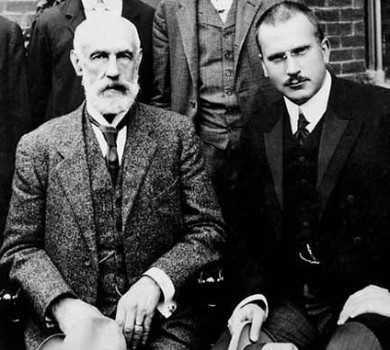 The pioneering ideas of Sigmund Freud (above left) and Carl Gustav Jung (right), combined with the developments
in neurosugery during the last century have made us aware of the
powerful electrophysiological processes that influence behaviour. The
introduction of a wide range of drugs to manage mood and behaviour, that modify the complex network of chemical pathways in the brain, continue to be investigated and tested.
The pioneering ideas of Sigmund Freud (above left) and Carl Gustav Jung (right), combined with the developments
in neurosugery during the last century have made us aware of the
powerful electrophysiological processes that influence behaviour. The
introduction of a wide range of drugs to manage mood and behaviour, that modify the complex network of chemical pathways in the brain, continue to be investigated and tested.
 Perhaps the most familiar aspect of brain related research in the News regularly is dementia and Alzheimer's disease. Like cancer, these brain related disorders have a wide range of causes, that we are only just beginning to get to grips with.The deterioration of tissue in specific regions of the brain can result in some very strange behaviours, not only memory loss. My own father suffered from Lewy Body dementia, which was characterised by halucinations of exotic animals1 When this was first explained to me some years ago, I was skeptical.
But after a brief dig around the medical literature and several
experiences with my dad, I was convinced. I still find these phenomena
very difficult to understand, but experiences or images that we are exposed
to both in the real world and on a screen, can clearly be retained and
re-formatted into "dream like" sequences whilst conscious. We have a
long way to go to understand such complex phenomena, but I
am confident that as we go on this journey in Science and Medicine, the
processes will unfold, and new therapies will be found to "fix" such
problems and go some way to restoring "quality of life" in the elderly (and actually in some relatively young adults too!).
Perhaps the most familiar aspect of brain related research in the News regularly is dementia and Alzheimer's disease. Like cancer, these brain related disorders have a wide range of causes, that we are only just beginning to get to grips with.The deterioration of tissue in specific regions of the brain can result in some very strange behaviours, not only memory loss. My own father suffered from Lewy Body dementia, which was characterised by halucinations of exotic animals1 When this was first explained to me some years ago, I was skeptical.
But after a brief dig around the medical literature and several
experiences with my dad, I was convinced. I still find these phenomena
very difficult to understand, but experiences or images that we are exposed
to both in the real world and on a screen, can clearly be retained and
re-formatted into "dream like" sequences whilst conscious. We have a
long way to go to understand such complex phenomena, but I
am confident that as we go on this journey in Science and Medicine, the
processes will unfold, and new therapies will be found to "fix" such
problems and go some way to restoring "quality of life" in the elderly (and actually in some relatively young adults too!).In Part 2, I will take a look at the blood supply and the nerves that link the body and brain and in the final part, I shall look at the way the brain interprets the senses: sight, smell, sound, taste and touch. Let me have your views in the comments box below!