When
I think of car tyres, I don't automatically think of Biological
molecules, but latex is the product of an unusual biosynthetic pathway
in the rubber plant Hevea brasiliensis, the Pará rubber tree, sharinga
tree, or, most commonly, simply, the rubber tree (shown on the LHS).
Isopentenyl pyrophosphate is the monomeric precursor (above RHS) for the
biosynthesis of the rubber polymer. The industrial process for
generating synthetic rubber (SR) follows a quite different route, but
the polymeric product is essentially the same. In addition, each natural
rubber polymer (NR) varies in "formulation" since the rubber particles
are associated with lipids and proteins that are species and
cell-specific. I won't have time to discuss this here, but you can read
more about this fascinating natural product here). The polymeric backbone is shown below.
When
I think of car tyres, I don't automatically think of Biological
molecules, but latex is the product of an unusual biosynthetic pathway
in the rubber plant Hevea brasiliensis, the Pará rubber tree, sharinga
tree, or, most commonly, simply, the rubber tree (shown on the LHS).
Isopentenyl pyrophosphate is the monomeric precursor (above RHS) for the
biosynthesis of the rubber polymer. The industrial process for
generating synthetic rubber (SR) follows a quite different route, but
the polymeric product is essentially the same. In addition, each natural
rubber polymer (NR) varies in "formulation" since the rubber particles
are associated with lipids and proteins that are species and
cell-specific. I won't have time to discuss this here, but you can read
more about this fascinating natural product here). The polymeric backbone is shown below.

The
starting point for rubber biosynthesis is IPP (half of the molecule of
the month: the second half is its polymerised form, rubber). The general
pathway for rubber biosynthesis from IPP is shown below). You can read a
recent genomic analysis of the pathways here.
What most interests me about rubber is the idea that cells can make such apparently "Inert", flexible insulating molecules which are in some ways the opposite of our own bones or a snail's shell.
The properties of rubber have led to a major industry and the development of industrial processes that convert NR into a useful product, such as in the manufacture of car and bicycle tyres. The
process you will all have heard of is vulcanisation. Essentially, this
proces adds sulphur to rubber (although other compounds may be added),
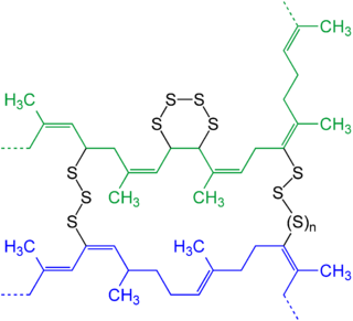
leading to a more robust, stiffer form of the polymer (see RHS). Vulcanisation takes its name from Vulcan, the Roman god
of fire and was patented most successfully by an American amateur
chemist, Charles Goodyear (the English industrial chemist Thomas Hancock also filed a patent on the method of vulcanisation pioneer, but this is another story). Like natural collagen, which forms spontaneous triple helices followed by age related cross links, rubber can be converted into a
whole range of formulations in order to meet the requirements of a
particular application. I am also reminded of disulphide linkages that
link the two chains of insulin together, or the heavy and light chains of antibody molecules. The drive to find ways of modifying the elastic and mechanical properties of rubber have led to a solution that Nature also adopts (or should I say adopted, a little earlier). If you find this area interesting, you may like to look up elastin, a polypeptide (family) with a repeated sequence of the type: GVPGXG (where X cannot be P).
 Economically, the international demand for rubber as an essential raw material is on the increase. Already, global consumption is nearly 11 million metric tons each year,
and demand is rapidly increasing. The USA is currently totally
dependent upon the import of more than 1.2 million metric tons of NR per year from tropically grown sources. The USA's rubber products trade is worth over $18
billion annually. For more than a century, Hevea brasiliensis has provided almost all of the world's natural rubber,
from a plantation and smallholding system involving tapping latex by
hand. In biological terms, latex production is thought to bring evolutionary benefits such as plant defence, and while Hevea is the main source of NR, many plants produce latex. There is no doubt that as a natural product, rubber is one of the most valuable and widely utilised non-enzymatic Bio-polymers. However, as synthetic biologists are beginning to successfully target complex metabolic pathways, such as the antimalarial secondary metabolite Artemisinin, rubber is also very much on their radar. Once again the ethics of destroying a major economy of a developing part of the world with GMOs will undoubtedly hit the headlines at some point. We need to have more discussions around such challenges.
Economically, the international demand for rubber as an essential raw material is on the increase. Already, global consumption is nearly 11 million metric tons each year,
and demand is rapidly increasing. The USA is currently totally
dependent upon the import of more than 1.2 million metric tons of NR per year from tropically grown sources. The USA's rubber products trade is worth over $18
billion annually. For more than a century, Hevea brasiliensis has provided almost all of the world's natural rubber,
from a plantation and smallholding system involving tapping latex by
hand. In biological terms, latex production is thought to bring evolutionary benefits such as plant defence, and while Hevea is the main source of NR, many plants produce latex. There is no doubt that as a natural product, rubber is one of the most valuable and widely utilised non-enzymatic Bio-polymers. However, as synthetic biologists are beginning to successfully target complex metabolic pathways, such as the antimalarial secondary metabolite Artemisinin, rubber is also very much on their radar. Once again the ethics of destroying a major economy of a developing part of the world with GMOs will undoubtedly hit the headlines at some point. We need to have more discussions around such challenges.
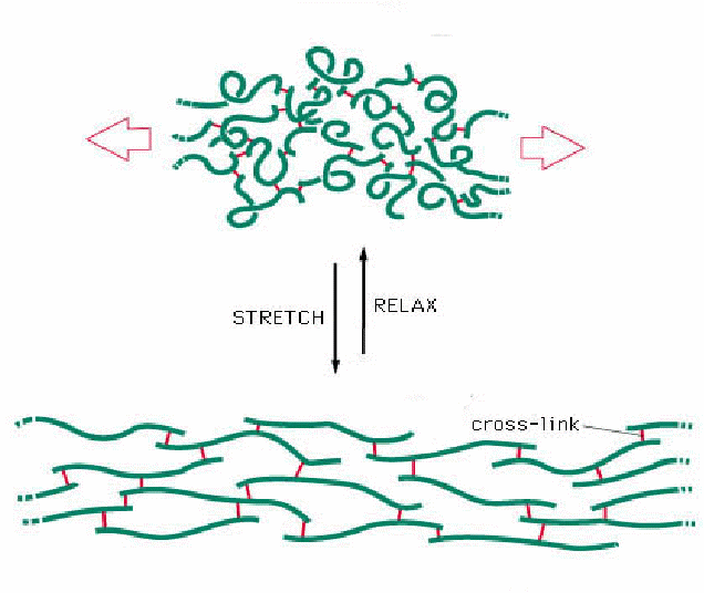 As a final point for discussion in a tutorial, one of the most familiar properties of rubber polymers is their elasticity: i.e. rubber can be reversibly stretched. When you stretch an elastic band it gets hot, when you let go, the rubber cools. The "ground state", or unstretched polymer chains, when relaxed are disordered (recall the second law of thermodynamics), however as they are stretched, the polymer chains align, becoming more organised, relative to the unstretched, ground state, thereby reducing entropy. As a result, heat is released. Stretching requires a physical input of energy and is a relatively slow process: snapping back to the ground state, on the other hand, is spontaneous. In kinetic terms, this provides a great example of the "power" of entropy, oh and it also provides food for kinetic thought too! Finally, the the sustainable and reversible elasticity of rubber is improved by vulcanisation: can this help us understand and explain the role of disulphide bonds in protein structure? These phenomena are encountered frequently when
biophysicists explore the thermodynamics and kinetics of protein
folding. I think a few elastic bands are a simple way to seed a tutorial
on the kinetics and thermodynamics of macromolecular folding? A final point for discussion could be the observation that rubber bands typically "perish" over a year or so. Why? This maybe more useful in discussions of nucleic acid polymers? Anyway, I hope this Molecule of the Month illustrates the Venn Diagram between Life Sciences. Chemistry and Physics!
As a final point for discussion in a tutorial, one of the most familiar properties of rubber polymers is their elasticity: i.e. rubber can be reversibly stretched. When you stretch an elastic band it gets hot, when you let go, the rubber cools. The "ground state", or unstretched polymer chains, when relaxed are disordered (recall the second law of thermodynamics), however as they are stretched, the polymer chains align, becoming more organised, relative to the unstretched, ground state, thereby reducing entropy. As a result, heat is released. Stretching requires a physical input of energy and is a relatively slow process: snapping back to the ground state, on the other hand, is spontaneous. In kinetic terms, this provides a great example of the "power" of entropy, oh and it also provides food for kinetic thought too! Finally, the the sustainable and reversible elasticity of rubber is improved by vulcanisation: can this help us understand and explain the role of disulphide bonds in protein structure? These phenomena are encountered frequently when
biophysicists explore the thermodynamics and kinetics of protein
folding. I think a few elastic bands are a simple way to seed a tutorial
on the kinetics and thermodynamics of macromolecular folding? A final point for discussion could be the observation that rubber bands typically "perish" over a year or so. Why? This maybe more useful in discussions of nucleic acid polymers? Anyway, I hope this Molecule of the Month illustrates the Venn Diagram between Life Sciences. Chemistry and Physics!
August
is usually the hottest month in the Northern Hemisphere and a time when
our skin can be exposed to risk from both high temperatures and ultra
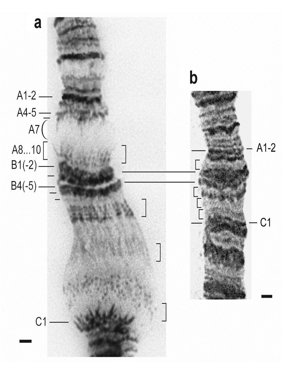
violet irradiation, it seemed timely then to pick a class of molecule that helps us deal with "heat shock". The "puffing" of chromatin (compare a and b left from Michael Ashburner's work) is associated with the "switching on " of heat shock genes. This is a Biological phenomenon that was described over 50 years ago in (mainly) Drosophila, and later captured the interest of an old friend of mine who sadly passed away last month, Tony (AR) Clarke.
It is also a molecule (well a class of molecule really) whose structure
and function has been the subject of Biochemical and Biophysical
investigation in a number of other UK labs, including Laurence Pearl's lab at Sussex and at the Martinsried Max Planck Institute in the laboratory of Ulrich Hartl. And since the publication of the first electron microscopy images and subsequent crystal structures, the number of molecular studies of this class of proteins has increased significantly.
In an earlier post, I discussed the fundamental significance of Christian B Anfinsen's work on the relationship between primary and tertiary structure in proteins. You may also recall (or take a look?) the caveats emerging, that suggest a given primary structure does not always give rise to a unique tertiary structure. However, there is another important mechanism involved in the translation (I use this word in its general sense) of protein sequence into three dimensional structure and function, and it has its origins in the work on "heat shock". It is now time to introduce the concept of the "molecular chaperone". The word chaperone was formerly used in the context of social norms in relation to the "appropriateness" of a single women to be "out" in public. A chaperone would, in this context, be typically an older woman (maybe an aunt or family friend) who would accompany a young, single woman to social events or visits to Fortnum and Masons, for a little retail therapy. The word is derived from old French, meaning a cover or hood (un chapeau: a hat, used to be in the first division of French vocabulary lists when I was at school). In some ways these two function: a covering and a protector,
describe the properties of chaperone proteins very nicely. These
molecules provide a safe haven for some newly synthesised proteins (see
the image from Ulrich Hartl's web site above), where the formation of tertiary structure requires a "leg up". I think this wiki site provides a nice, simple introduction to chaperone proteins. More detailed open access reviews include this one from Laurence Pearl in which he discusses Hsp90 structure and function in some detail, and refers extensively to the concept of "client" proteins, serviced by this particular molecular chaperone. I should also add that molecular chaperones are also referred to as chaperonins, but I think we can handle that!
Before I begin. Think for a moment about how you might "design" a molecular chaperone. This could form the basis of a nice "brainstorming" tutorial? Does every protein coding gene require its own chaperone to assist in folding, or perhaps more accurately prevent inappropriate aggregation: the primary role of chaperones in heat shock? How diverse are proteins in terms of molecular volume? Could a series of molecular "cages" say, accommodate small, medium and large proteins? How would a chaperone "attract" an unfolded, or "wayward" polypeptide chain and provide it with a safe haven? What is it that the safe haven could offer that the cytosol does not? How does the chaperone know when a protein is folded correctly? And when to let it go? How long does it take a non-Anfinsen polypeptide to emerge from a chaperone? What proportion of a cellular proteome is chaperone dependent? These and other considerations must be addressed in "designing" such a protein. I think this is a good undergraduate tutorial challenge.

The
structure on the left is a complex between the cylindrical GroEL and
the cap protein, GroES, one of the first chaperones to be described in
biochemical detail. The "channel" of GroEL is approximately 5nm, which can accommodate some, but not all polypeptides. However, if we consider its modus operandi, we can begin to appreciate that the mouth of the chaperone only needs to accommodate the unfolded (or partially folded) polypeptide chain. It is therefore an elongated cylinder and its interior attenuates the hydrophobic effect that drives many proteins to fold in the Anfinsen way. (The dimensions are given in the diagram below in Angstrom units: divide by 10 to covert to nm).
 Clearly, every polypeptide chain is different, and yet there are only a handful of chaperones (see Laurence Pearl's review for a discussion of co-chaperones and clients), chaperones must have been selected to be shared between several thousand unique ORFs (in E.coli): there must be some general interactions that offer an alternative to those available in the cellular milieu. Remember in the Anfinsen model a polypeptide chain is effectively "immersed" in water and immediately self-organises through backbone and side chain interactions,
driven by the partitioning of polar and non-polar groups. The fine
tuning of the three dimensional structure comes through the directional stabilisation of hydrogen bonds, ionic interactions and equilibration of hydrophobic interactions to achieve (mostly) a unique, minimum free energy state.
For non-Anfinsen polypeptides, a little help is needed to keep the polypeptide chain along the "straight and narrow", by closing down alternative, non-productive folding routes and inappropriate inter-molecular liaisons (such as those arising through aggregation). The diagram below provides an indication of the stages in in chaperone
mediated protein folding, which as you can see is energy requiring. It
is therefore important to recognise that this process is a necessary
evil, rather than a luxury item, since the correct assembly of several complex protein functions requires assistance. There must therefore be a co-dependence of the evolution of such proteins and their chaperone partners. The client: chaperone relationship, is itself the subject of much interest in the literature and is being exploited in the search for new therapeutic molecules (see work from the Pearl lab, for example).
Clearly, every polypeptide chain is different, and yet there are only a handful of chaperones (see Laurence Pearl's review for a discussion of co-chaperones and clients), chaperones must have been selected to be shared between several thousand unique ORFs (in E.coli): there must be some general interactions that offer an alternative to those available in the cellular milieu. Remember in the Anfinsen model a polypeptide chain is effectively "immersed" in water and immediately self-organises through backbone and side chain interactions,
driven by the partitioning of polar and non-polar groups. The fine
tuning of the three dimensional structure comes through the directional stabilisation of hydrogen bonds, ionic interactions and equilibration of hydrophobic interactions to achieve (mostly) a unique, minimum free energy state.
For non-Anfinsen polypeptides, a little help is needed to keep the polypeptide chain along the "straight and narrow", by closing down alternative, non-productive folding routes and inappropriate inter-molecular liaisons (such as those arising through aggregation). The diagram below provides an indication of the stages in in chaperone
mediated protein folding, which as you can see is energy requiring. It
is therefore important to recognise that this process is a necessary
evil, rather than a luxury item, since the correct assembly of several complex protein functions requires assistance. There must therefore be a co-dependence of the evolution of such proteins and their chaperone partners. The client: chaperone relationship, is itself the subject of much interest in the literature and is being exploited in the search for new therapeutic molecules (see work from the Pearl lab, for example).
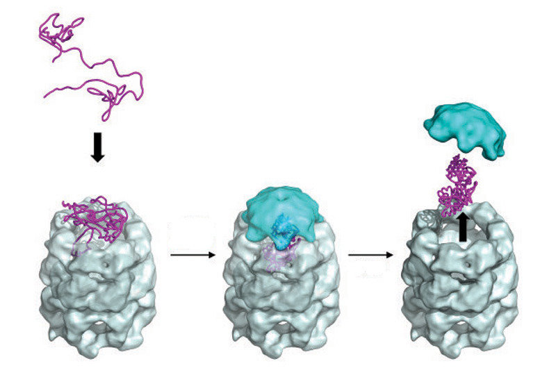
 The figure on the LHS, is taken from a review by Bigotti and Clarke and described the stages in the "capture" of a polypeptide substrate by the GroE system. Gro EL is in grey and blue, with the polypeptide sandwiched between the "mouthparts" which pull in the polypetide substrate in response to ATP addition. In this form, GroES now
binds rapidly with high affinity and the polypeptide enters the cavity
where folding takes place. The release of the protein follows two possible paths in vivo. In the "standard" sense, the folded protein is released for duty. However, unfolded polypeptides can also be sequestered for degradation via the proteosome in a similar, but opposite way. It must be remembered that a cellular proteome represents a steady state of synthesis and turn-over and similarly, all enzymatic reactions are in principle reversible: directionality is imposed by the downstream, biochemical demands.
Returning to the idea of a brainstorming turorial, I always think it is a good exercise to ask students to work out the phenomenal kinetic challenge of cell division, starting from an unreplicated and unexpressed genome comprising 3 000 genes and sufficient glucose to t urn a single colony inoculum into say 5g cells in a litre culture overnight! It is then that you begin to appreciate just how successful evolution has been! What I have never done is to compare the time taken for a an Anfinsen and a non-Anfinsen protein of the same size to "report for duty". Time to read the literature more thoroughly, I think.
The figure on the LHS, is taken from a review by Bigotti and Clarke and described the stages in the "capture" of a polypeptide substrate by the GroE system. Gro EL is in grey and blue, with the polypeptide sandwiched between the "mouthparts" which pull in the polypetide substrate in response to ATP addition. In this form, GroES now
binds rapidly with high affinity and the polypeptide enters the cavity
where folding takes place. The release of the protein follows two possible paths in vivo. In the "standard" sense, the folded protein is released for duty. However, unfolded polypeptides can also be sequestered for degradation via the proteosome in a similar, but opposite way. It must be remembered that a cellular proteome represents a steady state of synthesis and turn-over and similarly, all enzymatic reactions are in principle reversible: directionality is imposed by the downstream, biochemical demands.
Returning to the idea of a brainstorming turorial, I always think it is a good exercise to ask students to work out the phenomenal kinetic challenge of cell division, starting from an unreplicated and unexpressed genome comprising 3 000 genes and sufficient glucose to t urn a single colony inoculum into say 5g cells in a litre culture overnight! It is then that you begin to appreciate just how successful evolution has been! What I have never done is to compare the time taken for a an Anfinsen and a non-Anfinsen protein of the same size to "report for duty". Time to read the literature more thoroughly, I think.