August
is usually the hottest month in the Northern Hemisphere and a time when
our skin can be exposed to risk from both high temperatures and ultra
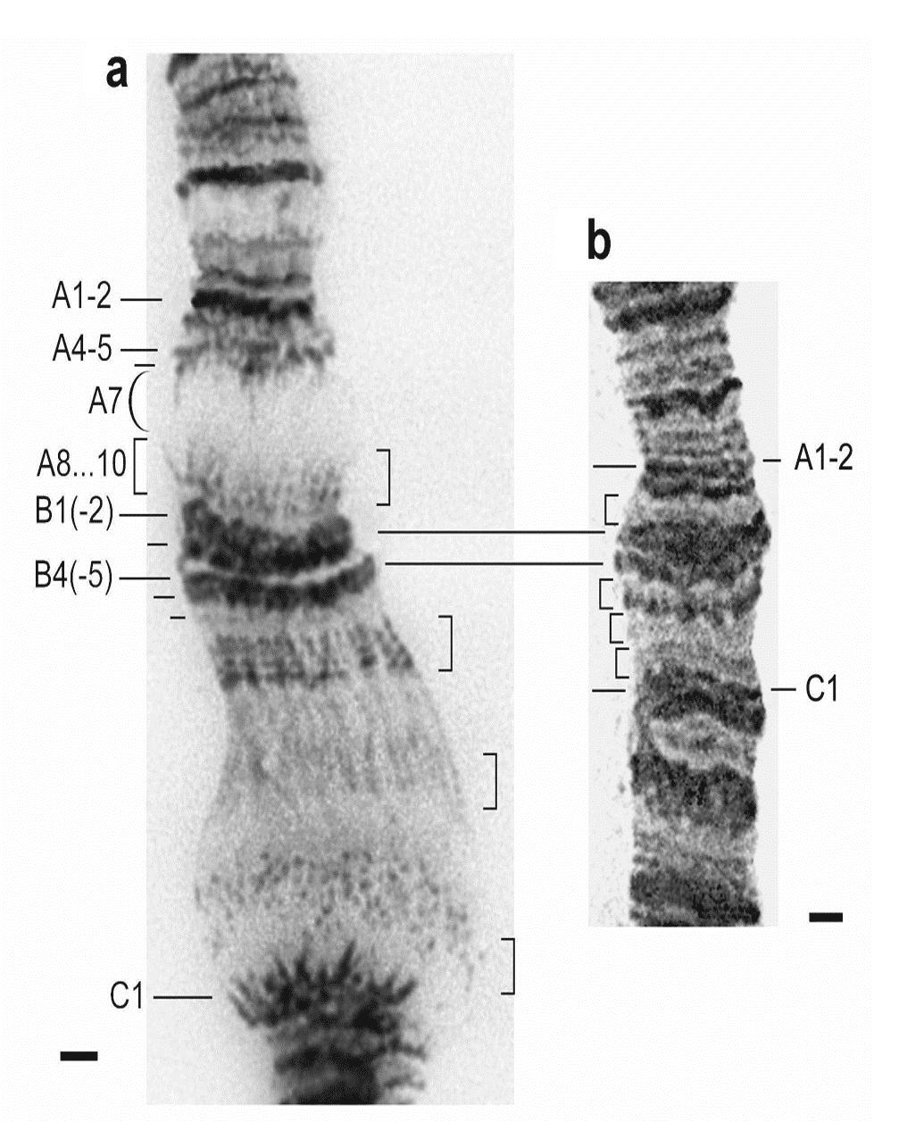
violet irradiation, it seemed timely then to pick a class of molecule that helps us deal with "heat shock". The "puffing" of chromatin (compare a and b left from Michael Ashburner's work) is associated with the "switching on " of heat shock genes. This is a Biological phenomenon that was described over 50 years ago in (mainly) Drosophila, and later captured the interest of an old friend of mine who sadly passed away last month, Tony (AR) Clarke.
It is also a molecule (well a class of molecule really) whose structure
and function has been the subject of Biochemical and Biophysical
investigation in a number of other UK labs, including Laurence Pearl's lab at Sussex and at the Martinsried Max Planck Institute in the laboratory of Ulrich Hartl. And since the publication of the first electron microscopy images and subsequent crystal structures, the number of molecular studies of this class of proteins has increased significantly.
In an earlier post, I discussed the fundamental significance of Christian B Anfinsen's work on the relationship between primary and tertiary structure in proteins. You may also recall (or take a look?) the caveats emerging, that suggest a given primary structure does not always give rise to a unique tertiary structure. However, there is another important mechanism involved in the translation (I use this word in its general sense) of protein sequence into three dimensional structure and function, and it has its origins in the work on "heat shock". It is now time to introduce the concept of the "molecular chaperone". The word chaperone was formerly used in the context of social norms in relation to the "appropriateness" of a single women to be "out" in public. A chaperone would, in this context, be typically an older woman (maybe an aunt or family friend) who would accompany a young, single woman to social events or visits to Fortnum and Masons, for a little retail therapy. The word is derived from old French, meaning a cover or hood (un chapeau: a hat, used to be in the first division of French vocabulary lists when I was at school). In some ways these two function: a covering and a protector,
describe the properties of chaperone proteins very nicely. These
molecules provide a safe haven for some newly synthesised proteins (see
the image from Ulrich Hartl's web site above), where the formation of tertiary structure requires a "leg up". I think this wiki site provides a nice, simple introduction to chaperone proteins. More detailed open access reviews include this one from Laurence Pearl in which he discusses Hsp90 structure and function in some detail, and refers extensively to the concept of "client" proteins, serviced by this particular molecular chaperone. I should also add that molecular chaperones are also referred to as chaperonins, but I think we can handle that!
Before I begin. Think for a moment about how you might "design" a molecular chaperone. This could form the basis of a nice "brainstorming" tutorial? Does every protein coding gene require its own chaperone to assist in folding, or perhaps more accurately prevent inappropriate aggregation: the primary role of chaperones in heat shock? How diverse are proteins in terms of molecular volume? Could a series of molecular "cages" say, accommodate small, medium and large proteins? How would a chaperone "attract" an unfolded, or "wayward" polypeptide chain and provide it with a safe haven? What is it that the safe haven could offer that the cytosol does not? How does the chaperone know when a protein is folded correctly? And when to let it go? How long does it take a non-Anfinsen polypeptide to emerge from a chaperone? What proportion of a cellular proteome is chaperone dependent? These and other considerations must be addressed in "designing" such a protein. I think this is a good undergraduate tutorial challenge.

The
structure on the left is a complex between the cylindrical GroEL and
the cap protein, GroES, one of the first chaperones to be described in
biochemical detail. The "channel" of GroEL is approximately 5nm, which can accommodate some, but not all polypeptides. However, if we consider its modus operandi, we can begin to appreciate that the mouth of the chaperone only needs to accommodate the unfolded (or partially folded) polypeptide chain. It is therefore an elongated cylinder and its interior attenuates the hydrophobic effect that drives many proteins to fold in the Anfinsen way. (The dimensions are given in the diagram below in Angstrom units: divide by 10 to covert to nm).
 Clearly, every polypeptide chain is different, and yet there are only a handful of chaperones (see Laurence Pearl's review for a discussion of co-chaperones and clients), chaperones must have been selected to be shared between several thousand unique ORFs (in E.coli): there must be some general interactions that offer an alternative to those available in the cellular milieu. Remember in the Anfinsen model a polypeptide chain is effectively "immersed" in water and immediately self-organises through backbone and side chain interactions,
driven by the partitioning of polar and non-polar groups. The fine
tuning of the three dimensional structure comes through the directional stabilisation of hydrogen bonds, ionic interactions and equilibration of hydrophobic interactions to achieve (mostly) a unique, minimum free energy state.
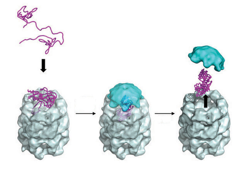
For non-Anfinsen polypeptides, a little help is needed to keep the polypeptide chain along the "straight and narrow", by closing down alternative, non-productive folding routes and inappropriate inter-molecular liaisons (such as those arising through aggregation). The diagram below provides an indication of the stages in in chaperone
mediated protein folding, which as you can see is energy requiring. It
is therefore important to recognise that this process is a necessary
evil, rather than a luxury item, since the correct assembly of several complex protein functions requires assistance. There must therefore be a co-dependence of the evolution of such proteins and their chaperone partners. The client: chaperone relationship, is itself the subject of much interest in the literature and is being exploited in the search for new therapeutic molecules (see work from the Pearl lab, for example).
Clearly, every polypeptide chain is different, and yet there are only a handful of chaperones (see Laurence Pearl's review for a discussion of co-chaperones and clients), chaperones must have been selected to be shared between several thousand unique ORFs (in E.coli): there must be some general interactions that offer an alternative to those available in the cellular milieu. Remember in the Anfinsen model a polypeptide chain is effectively "immersed" in water and immediately self-organises through backbone and side chain interactions,
driven by the partitioning of polar and non-polar groups. The fine
tuning of the three dimensional structure comes through the directional stabilisation of hydrogen bonds, ionic interactions and equilibration of hydrophobic interactions to achieve (mostly) a unique, minimum free energy state.
For non-Anfinsen polypeptides, a little help is needed to keep the polypeptide chain along the "straight and narrow", by closing down alternative, non-productive folding routes and inappropriate inter-molecular liaisons (such as those arising through aggregation). The diagram below provides an indication of the stages in in chaperone
mediated protein folding, which as you can see is energy requiring. It
is therefore important to recognise that this process is a necessary
evil, rather than a luxury item, since the correct assembly of several complex protein functions requires assistance. There must therefore be a co-dependence of the evolution of such proteins and their chaperone partners. The client: chaperone relationship, is itself the subject of much interest in the literature and is being exploited in the search for new therapeutic molecules (see work from the Pearl lab, for example).
 The figure on the LHS, is taken from a review by Bigotti and Clarke and described the stages in the "capture" of a polypeptide substrate by the GroE system. Gro EL is in grey and blue, with the polypeptide sandwiched between the "mouthparts" which pull in the polypetide substrate in response to ATP addition. In this form, GroES now
binds rapidly with high affinity and the polypeptide enters the cavity
where folding takes place. The release of the protein follows two possible paths in vivo. In the "standard" sense, the folded protein is released for duty. However, unfolded polypeptides can also be sequestered for degradation via the proteosome in a similar, but opposite way. It must be remembered that a cellular proteome represents a steady state of synthesis and turn-over and similarly, all enzymatic reactions are in principle reversible: directionality is imposed by the downstream, biochemical demands.
Returning to the idea of a brainstorming turorial, I always think it is a good exercise to ask students to work out the phenomenal kinetic challenge of cell division, starting from an unreplicated and unexpressed genome comprising 3 000 genes and sufficient glucose to t urn a single colony inoculum into say 5g cells in a litre culture overnight! It is then that you begin to appreciate just how successful evolution has been! What I have never done is to compare the time taken for a an Anfinsen and a non-Anfinsen protein of the same size to "report for duty". Time to read the literature more thoroughly, I think.
The figure on the LHS, is taken from a review by Bigotti and Clarke and described the stages in the "capture" of a polypeptide substrate by the GroE system. Gro EL is in grey and blue, with the polypeptide sandwiched between the "mouthparts" which pull in the polypetide substrate in response to ATP addition. In this form, GroES now
binds rapidly with high affinity and the polypeptide enters the cavity
where folding takes place. The release of the protein follows two possible paths in vivo. In the "standard" sense, the folded protein is released for duty. However, unfolded polypeptides can also be sequestered for degradation via the proteosome in a similar, but opposite way. It must be remembered that a cellular proteome represents a steady state of synthesis and turn-over and similarly, all enzymatic reactions are in principle reversible: directionality is imposed by the downstream, biochemical demands.
Returning to the idea of a brainstorming turorial, I always think it is a good exercise to ask students to work out the phenomenal kinetic challenge of cell division, starting from an unreplicated and unexpressed genome comprising 3 000 genes and sufficient glucose to t urn a single colony inoculum into say 5g cells in a litre culture overnight! It is then that you begin to appreciate just how successful evolution has been! What I have never done is to compare the time taken for a an Anfinsen and a non-Anfinsen protein of the same size to "report for duty". Time to read the literature more thoroughly, I think.
 Bacteriophage, or more commonly 'phage for short, is the name given to viruses that infect bacteria (you can see the schematic diagram of phage T4 on the left). You will have come across the suffix phage in oesophagus or as a prefix in Phagocytosis: it has Greek origins and means to eat. I first came across bacteriophage in 1977 during an
undergraduate lecture by Dr. Bernard Fry at Sheffield. I was
immediately hooked! Here was one of the first scientific conundrums I
faced: are bacteriophage dead or alive? Is a biological "system" that requires the "machinery" of a bacterial host to replicate (phage,
incidentally outnumber most species on the planet), a living organism
or an inert molecule? For me it sparked an interest in the origin of
life, and it
also sparked another interest, in the area molecular structure and
function. Our appetites (well those of us who found these lectures
fascinating) were whetted further by what felt at the time to be
powerful Electron Microscopy (EM) images. Fifty years on from these classic experiments, in the light of the marvellous structural biology images of multi-subunit enzymes such as the ribosome, with which we have become so familiar; the early EMs appear, well, maybe a little dated?
Bacteriophage, or more commonly 'phage for short, is the name given to viruses that infect bacteria (you can see the schematic diagram of phage T4 on the left). You will have come across the suffix phage in oesophagus or as a prefix in Phagocytosis: it has Greek origins and means to eat. I first came across bacteriophage in 1977 during an
undergraduate lecture by Dr. Bernard Fry at Sheffield. I was
immediately hooked! Here was one of the first scientific conundrums I
faced: are bacteriophage dead or alive? Is a biological "system" that requires the "machinery" of a bacterial host to replicate (phage,
incidentally outnumber most species on the planet), a living organism
or an inert molecule? For me it sparked an interest in the origin of
life, and it
also sparked another interest, in the area molecular structure and
function. Our appetites (well those of us who found these lectures
fascinating) were whetted further by what felt at the time to be
powerful Electron Microscopy (EM) images. Fifty years on from these classic experiments, in the light of the marvellous structural biology images of multi-subunit enzymes such as the ribosome, with which we have become so familiar; the early EMs appear, well, maybe a little dated?
 As me and my classmates passed through the various Biochemistry lecture
courses in metabolism and enzymology, we finally caught up with these
strange hybrids between living organisms and molecular assemblies in
crystallography lectures, when Professor Pauline Harrison showed us
images of the plant virus: Tobacco Mosaic Virus (RHS). It was clear that
phage and viruses would hold a beauty that just needed time and patience, and the development of better methods of sample preparation, data collection and analysis, for high resolution structures to emerge. Well those methods have
finally arrived and the technique of cryo EM is now set to provide us
with molecular details of these macromolecular machines. I shall provide a small amount of background information to help provide a context to the work.
As me and my classmates passed through the various Biochemistry lecture
courses in metabolism and enzymology, we finally caught up with these
strange hybrids between living organisms and molecular assemblies in
crystallography lectures, when Professor Pauline Harrison showed us
images of the plant virus: Tobacco Mosaic Virus (RHS). It was clear that
phage and viruses would hold a beauty that just needed time and patience, and the development of better methods of sample preparation, data collection and analysis, for high resolution structures to emerge. Well those methods have
finally arrived and the technique of cryo EM is now set to provide us
with molecular details of these macromolecular machines. I shall provide a small amount of background information to help provide a context to the work.
 The mechanism of phage infection differs from the viral infection of eukaryotic cells, since the latter are generally free from a peptidoglycan layer.
Phage such as T4 (one of the T-even phages) make an initial interaction
with a surface molecule (part of the peptidoglycan wall), which is followed by the release of the "landing gear", or the tail fibres and base plate. [The nomenclature of phage is a little idiosyncratic, but the
T even phage are collectively more complex in structure than the T odd
phage (T5, T7 etc): the T odd group do not have a contractile sheath,
but rather a tail]. I wont say much more about the T odd phages here.
Eukaryotic viruses generally make similar interactions, but then, owing
to the similarity of the viral "coat" to a plasma membrane, a fusion event takes place. However, I won't say anything further about viruses, either, for now. T4 phage all possess a geometrically symmetrical head or capsid, in which around 170 000 base pairs of genomic DNA are packaged. The focus of this post is the base plate, sheath and landing gear which surround the "syringe" like structure, used by T4 to inject its genomic payload into its chosen host. An incredible piece of nano-architecture!
The mechanism of phage infection differs from the viral infection of eukaryotic cells, since the latter are generally free from a peptidoglycan layer.
Phage such as T4 (one of the T-even phages) make an initial interaction
with a surface molecule (part of the peptidoglycan wall), which is followed by the release of the "landing gear", or the tail fibres and base plate. [The nomenclature of phage is a little idiosyncratic, but the
T even phage are collectively more complex in structure than the T odd
phage (T5, T7 etc): the T odd group do not have a contractile sheath,
but rather a tail]. I wont say much more about the T odd phages here.
Eukaryotic viruses generally make similar interactions, but then, owing
to the similarity of the viral "coat" to a plasma membrane, a fusion event takes place. However, I won't say anything further about viruses, either, for now. T4 phage all possess a geometrically symmetrical head or capsid, in which around 170 000 base pairs of genomic DNA are packaged. The focus of this post is the base plate, sheath and landing gear which surround the "syringe" like structure, used by T4 to inject its genomic payload into its chosen host. An incredible piece of nano-architecture!
A few words now about the methodology employed, since the
experimental methodology underpinning this work is itself a tour de
force. First the source material: the authors used a classic phage T4
mutant in order to persuade the host to mass produce the base-plate in the absence of the capsid,
drawing on a fine piece of bacterial genetics which provided phage
molecular biologists the opportunity to map assembly pathways. Take a
look at the lab web site of Jonathan King at MIT: still going strong after some remarkable work in the late '60s and early '70s. Taylor et al used a mutant phage that fails to complete assembly,
but instead leads to the accumulation of base plates in the host. From
such cells, it is possible to obtain mg quantities of the protein
complex, in a homogeneous form; ideal fro the downstream cryo -EM analysis. A superb example of the powerful combination of bacterial genetics and biochemistry.
 The development of the electron microscope in
Berlin in the late 1930s (the image RHS was taken in 1940), provided post-war physicists and biologists
with a powerful method for visualising viruses and bacteriophage in
particular. (Not forgetting the pioneering work
at the same time from George Palade and others on the ultra-structure
of mammalian and plant cells). As you can see above, the images reveal a clear attachment of numerous phage particles around the cell wall of the host with mature viruses either inside or laying flat on the surface of the host. However it wasn't until the 1990s that sample preparation combined with image processing methods made it possible to begin to see detail in the images.
The development of the electron microscope in
Berlin in the late 1930s (the image RHS was taken in 1940), provided post-war physicists and biologists
with a powerful method for visualising viruses and bacteriophage in
particular. (Not forgetting the pioneering work
at the same time from George Palade and others on the ultra-structure
of mammalian and plant cells). As you can see above, the images reveal a clear attachment of numerous phage particles around the cell wall of the host with mature viruses either inside or laying flat on the surface of the host. However it wasn't until the 1990s that sample preparation combined with image processing methods made it possible to begin to see detail in the images.
 It should be noted that X-ray crystallographers (using single crystals) had by the turn of this century, been successful in obtaining high resolution structures of "spherical" virus particles such as Tomato Bushy Stunt Virus. However, the last ten years has seen a gradual shift in technology, amongst structural biologists away from X-ray crystallography towards cryo-EM for the determination of multi-protein assemblies. As with all structure determination methods (including NMR), it will undoubtedly be the case that structural analysis and the interpretation of structure and function will benefit from the judicious choice of technique, depending on the question being asked. You can find a recent summary of cryo-EM technology here at the Baumeister Lab Home Page.
So, armed with a nice "production method" for base-plate particle and cryo-EM technology capable of resolving such particles at molecular resolution, Taylor et al have built on the work of Michael Rossmann's Lab and determined the structure of the T4 base plate, providing clues to the attachment and pre-injection stages of phage infection. The three stages:airborne (pre-attachment), approaching the target and landing (attachment) are intuitively clear from the structural models calculated from the electron density aided by published crystal structures of some of the individual components, and shown below (and in more detail here).
It should be noted that X-ray crystallographers (using single crystals) had by the turn of this century, been successful in obtaining high resolution structures of "spherical" virus particles such as Tomato Bushy Stunt Virus. However, the last ten years has seen a gradual shift in technology, amongst structural biologists away from X-ray crystallography towards cryo-EM for the determination of multi-protein assemblies. As with all structure determination methods (including NMR), it will undoubtedly be the case that structural analysis and the interpretation of structure and function will benefit from the judicious choice of technique, depending on the question being asked. You can find a recent summary of cryo-EM technology here at the Baumeister Lab Home Page.
So, armed with a nice "production method" for base-plate particle and cryo-EM technology capable of resolving such particles at molecular resolution, Taylor et al have built on the work of Michael Rossmann's Lab and determined the structure of the T4 base plate, providing clues to the attachment and pre-injection stages of phage infection. The three stages:airborne (pre-attachment), approaching the target and landing (attachment) are intuitively clear from the structural models calculated from the electron density aided by published crystal structures of some of the individual components, and shown below (and in more detail here).

As a Biochemist with a leaning towards Biophysics, the question that immediately presents itself to me is: how does folded (compact) conformation change to the extended landing conformation? Any protein synthesised in the cell (or
sometimes in the test-tube) folds (almost) spontaneously into its final
shape (see an earlier post on protein folding and ebola virus here).
Coalescence of hydrophobic side chains and subsequent coating of the
protein surface with polar side chains drives the formation of soluble
globular proteins. We also know that allosteric enzymes are poised in an
equilibrium between two conformational states (active and inactive) and
that small ligands can tip the equilibrium over to favour one of the two conformations. It seems to me that the forces that stabilise the folded legs are considerably weaker than those that persuade the legs to extend and clamp onto the surface of the appropriate host cell surface. (Imagine a set of legs that are jointed and ferromagnetic, stabilised in the folded state by the magnetic surface underneath the base-plate, being induced to unfold by a surface that is much more strongly magnetic: the bacterial cell wall).
 The final point I want to make relates to the extended structures that form the legs and the injection tube, or spike. I have captured an image from the protein data base of the small T4 protein gp5 (see RHS). If you look closely the spike is a cylinder formed from beta sheets. The spike ends in a tip that penetrates the bacterial cell and injects the DNA. What a fantastic example of structure and function in Nature at the molecular level. Similarly, the legs are extended structures which remind me of the extended helix in the calcium sensor protein calmodulin. Such structures are intrinsically unstable in the sense that they are vulnerable to proteases, however, when I see a crane on a large building site, I think of the engineering design that makes the tall structre sufficiently stable to move around heavy objects. Nature has achieved similar fetes at the molecular level....and this structure has many more features than I have touched on, so please read the article in Nature if you can access it, or go to Petr Leiman's web site here.

 Clearly, every polypeptide chain is different, and yet there are only a handful of chaperones (see Laurence Pearl's review for a discussion of co-chaperones and clients), chaperones must have been selected to be shared between several thousand unique ORFs (in E.coli): there must be some general interactions that offer an alternative to those available in the cellular milieu. Remember in the Anfinsen model a polypeptide chain is effectively "immersed" in water and immediately self-organises through backbone and side chain interactions,
driven by the partitioning of polar and non-polar groups. The fine
tuning of the three dimensional structure comes through the directional stabilisation of hydrogen bonds, ionic interactions and equilibration of hydrophobic interactions to achieve (mostly) a unique, minimum free energy state.
Clearly, every polypeptide chain is different, and yet there are only a handful of chaperones (see Laurence Pearl's review for a discussion of co-chaperones and clients), chaperones must have been selected to be shared between several thousand unique ORFs (in E.coli): there must be some general interactions that offer an alternative to those available in the cellular milieu. Remember in the Anfinsen model a polypeptide chain is effectively "immersed" in water and immediately self-organises through backbone and side chain interactions,
driven by the partitioning of polar and non-polar groups. The fine
tuning of the three dimensional structure comes through the directional stabilisation of hydrogen bonds, ionic interactions and equilibration of hydrophobic interactions to achieve (mostly) a unique, minimum free energy state.  The figure on the LHS, is taken from a review by Bigotti and Clarke and described the stages in the "capture" of a polypeptide substrate by the GroE system. Gro EL is in grey and blue, with the polypeptide sandwiched between the "mouthparts" which pull in the polypetide substrate in response to ATP addition. In this form, GroES now
binds rapidly with high affinity and the polypeptide enters the cavity
where folding takes place. The release of the protein follows two possible paths in vivo. In the "standard" sense, the folded protein is released for duty. However, unfolded polypeptides can also be sequestered for degradation via the proteosome in a similar, but opposite way. It must be remembered that a cellular proteome represents a steady state of synthesis and turn-over and similarly, all enzymatic reactions are in principle reversible: directionality is imposed by the downstream, biochemical demands.
The figure on the LHS, is taken from a review by Bigotti and Clarke and described the stages in the "capture" of a polypeptide substrate by the GroE system. Gro EL is in grey and blue, with the polypeptide sandwiched between the "mouthparts" which pull in the polypetide substrate in response to ATP addition. In this form, GroES now
binds rapidly with high affinity and the polypeptide enters the cavity
where folding takes place. The release of the protein follows two possible paths in vivo. In the "standard" sense, the folded protein is released for duty. However, unfolded polypeptides can also be sequestered for degradation via the proteosome in a similar, but opposite way. It must be remembered that a cellular proteome represents a steady state of synthesis and turn-over and similarly, all enzymatic reactions are in principle reversible: directionality is imposed by the downstream, biochemical demands.


